Fig. 14.1
Noncarious cervical lesions can range from shallow depressions that involve enamel only (a), enamel and radicular tooth structure (b, c), or radicular tooth structure only. They vary in shape based on the combination of etiologic factors present. Lesions can progress to a size that can compromise tooth vitality (d)
Although evidence of occlusal attrition and abrasion exists among the dentition of hunter-gather populations from thousands of years ago, the prevalence of noncarious cervical lesions has not been observed in remains of these populations. Noncarious cervical lesions should therefore be viewed as pathology unique to modern man [1]. Today, these lesions can be found in both children and adults. Prevalence studies have resulted in conflicting results. Shulman and Robinson recorded prevalence as low as 2 % [2], whereas Bergstrom and Eliasson [3] recorded findings of 90 %. In a review of 15 studies carried out between 1941 and 1991, Levitch et al. [4] reported the prevalence data for these lesions ranging between 5 and 85 %. The wide range can be explained in several ways. Some studies had small sample sizes and used very different age groups, and different studies classified lesions in different ways. When you add in the number of variables to consider within a population or within individuals of a population over time, it is not surprising that the prevalence data is so confusing.
Just as the prevalence of NCCLs is unclear, so is the exact picture of their intraoral distribution. Rees et al. [5] and Sognnaes et al. [6] reported that the lesions are most prevalent on the labial surface of maxillary incisors; whereas, Radentz et al. [7] reported maxillary first molars to be the most commonly affected teeth, and Zipkin and McLure [8] found the maxillary first premolars to be the most involved tooth. This variability in reported distribution patterns is possibly due to the confusing terminologies and variable diagnoses that were employed in different studies at different times. However, despite all of the flaws and contradictions of the early prevalence studies, some facts clearly emerge: the older the study population was, the greater the numbers of lesions per individual were found, and the larger the lesions were. Also, these lesions are almost exclusively found on the facial or buccal aspects of teeth, seldom on the lingual and rarely on the proximal surfaces.
14.2 Etiology of Noncarious Lesions
A number of theories regarding the etiological mechanism of tooth wear in the absence of caries have developed over time; however, the entire etiology of noncarious cervical lesions is still controversial. In 1907, Miller [9–11] conducted an exhaustive investigation of noncarious tooth surface lesions and was the first to associate these lesions, which he termed “wastings” with mechanical and chemical factors. NCCLs were thus first classified according to their supposed origin: abrasion or erosion. Abrasion was generally ascribed to either the toothbrush and or dentifrice pathologically wearing away tooth structure by mechanical or frictional forces; however, other factors that exert a repetitive and sometimes excessive force on teeth such as toothpicks, floss, removable appliances, and parafunctional habits can also contribute to cervical tooth wear (Fig. 14.2). Erosion has been defined as loss of tooth structure produced by chemical dissolution from acids other than those produced by bacteria [12]. In the strictest definition of the word, erosion is not a chemical mechanism but rather a physical mechanism by which wear is caused by friction from the movement of liquids. Therefore, a paradigm shift has been suggested to supplant the term “erosion” with the more accurate term “biocorrosion” defined as the chemical, biochemical, or electrochemical action that causes the molecular degradation of tooth substance [13]. Biocorrosion as a term takes into consideration more than just chemical exogenous and biochemical endogenous acids, it also encompasses proteolytic agents and the piezoelectric effects observed on dentin.
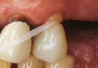
Fig. 14.2
Noncarious cervical lesions can result from aggressive or improperly implemented oral hygiene techniques. In this instance, the repetitive sawing action of the floss wore a notch in the root surface of the tooth (Photo courtesy of Dr. Stephan J Haney)
Another relatively new term to the discussion of NCCLs is that of “abfraction” introduced by Grippo in 1991 [14], and amended in 2004 [15]. Abfraction describes the microfracture of tooth substance in areas of stress concentration. The term abfraction has been abused and misused by the dental community as a generic designation for all NCCLs, whereas the etiology of these lesions is generally believed to be multifactorial. In order to appropriately address the causative factors surrounding noncarious cervical lesions in an individual, the clinician must appreciate the unique interplay among those factors for that individual. The three major potential mechanisms in lesion development consist of abrasion/friction, abfraction/stress, and biocorrosion. Failure to appropriately prevent and treat noncarious cervical lesions can result in their progression, the potential for tooth sensitivity, and, in severe cases, endodontic therapy or even tooth loss.
It is critical for a clinician to perform a thorough medical and dental history and to consider the complex interaction of the contributory factors and modifying factors before completing the diagnosis and initiating treatment. The following section will discuss these factors in more depth.
14.2.1 Biocorrosion
Biocorrosion of teeth can occur as a result of exogenous chemical and endogenous biochemical acids, as well as by proteolytic enzymes, and piezoelectric effects on dentin. The fact that tooth enamel and dentin can be dissolved by acid is a well-established fact, regardless of whether the acid is produced as a by-product of oral bacteria as is in the case of dental caries, ingested in the form of acid containing food or beverage, or intrinsically produced as in gastric reflux or vomiting or purging. Enamel is mainly composed of inorganic hydroxyapatite, with only a small percentage of organic matrix material, and, thus, it is readily disintegrated by acid. A pH below 5.5 has been shown to dissolve enamel [16]. Dentin contains less hydroxyapatite than enamel and more organic matrix material. Thus, the surface layer of the dentin hydroxyapatite can be demineralized by acid leaving the organic matrix, which is not water soluble but which is subject to attack by proteolytic enzymes (proteases) that can be produced by the microorganisms in plaque or come from gingival crevicular fluid. One of the reasons that gastroesophageal reflux disease or habitual regurgitation is so devastating to teeth is the combined exposure to the extremely acidic gastric juice and the proteolytic enzymes from the stomach (pepsin) and pancreas (trypsin).
It is interesting to note, when considering the effect that acids have on demineralizing tooth structure, that pH does not tell the whole story. Different acids have different corrosive potentials. Therefore, the pH of a substance alone is not totally predictive of its potential to cause biocorrosion. For acidic drinks, it was found that not only the pH value but also the type of acid, the amount of titratable acid (buffer capacity), and possibly chelating properties are factors in determining the progression of biocorrosion [17]. In vitro studies show that citric acid and phosphoric acid both produce more tissue loss than maleic acid [18]. The citrate ion may be particularly destructive because of its binding or chelating action on calcium. Larsen and Nyvad found that the addition of calcium and phosphate to acidic drinks reduced their ability to dissolve enamel [19]. Lussi et al. demonstrated the same protective effect of calcium and phosphate using the example of yogurt, which has a pH of close to 4 but has no corrosive effects on tooth structure because of its high calcium and phosphate content [17]. Additionally, the oral cavity has several protective mechanisms against biocorrosion, the most prominent being saliva. Salivary flow is increased by acid-induced stimulation of the glands. Clearance of acids from the oral cavity is, to a large extent, dependent on the saliva flow rate and the saliva buffering capacity. Low saliva flow rate and poor buffering capacity allow prolonged retention of extrinsic and intrinsic acids in the mouth, which will accelerate the biocorrosive process. Therefore, the quantity and the quality of a patient’s saliva is an important modifying factor to the progression of NCCLs and should be a part of patient evaluation. The pellicle on the tooth’s surface, which represents a diffusion barrier to the acids, also aids in preventing acids from directly dissolving the tooth mineral.
14.2.2 Abrasion
The loss of tooth structure in the cervical area of the tooth as a result of mechanical rubbing with some object is known as abrasion. This may be due to aggressive toothbrushing or flossing techniques, stiff brushes, the use of abrasive dentifrices, or even repetitive habits.
Normally, enamel is very resistant to wear, and abrasion is low especially as compared to dentin or cementum. However, the abrasion resistance of both enamel and dentin is weakened after exposure to acid. There is evidence that the loss of tooth structure from abrasion is accelerated when acid demineralization of enamel and dentin occurs prior to or during mechanical abrasion of those surfaces [20]. Fortunately, demineralized tooth structure can be remineralized if the weakened tooth structure is not removed as during brushing or other mechanical rubbing before it can be remineralized through exposure to saliva for a long enough period of time. In fact, when teeth are exposed to saliva, delaying brushing for as little as 1 h after an acid challenge can increase their resistance to abrasion [21]. The advice for patients to brush immediately after every meal needs to be re-evaluated and modified. Additionally, patients exhibiting NCCLs should be advised to use only a small amount of a minimally abrasive fluoride containing dentifrice with light force when brushing or skip the dentifrice all together and use a fluoride rinse instead.
Combining occlusal loading (as in abfraction) with abrasion does not seem to have the same additive effect resulting in tooth loss that combining abrasion with exposure to acid has. Litonjua et al. found that without any acids present, occlusal loading had no effect in creating NCCLs in recently extracted teeth subjected to toothpaste slurry and tooth brushing at the cervical margins. Brushing with the toothpaste slurry caused similar cervical wear patterns regardless of whether or not the tooth was loaded [22].
14.2.3 Abfraction
The theory that occlusal forces are an etiologic factor in the formation of NCCLs is a relatively new concept, and one that has definitely been the focus of much attention and controversy. The term “abfraction” which means “to break away” has been proposed for this phenomenon. Abfraction represents the mechanical flexure theory where tooth bending and flexing during function and parafunction create flexural stress in the cervical area of the tooth resulting in microfractures of the crystalline structure of the enamel and dentin in that area. The theory suggests that the lesion would continue to enlarge as the bending and flexing is repeated finally resulting in chipping away of the hard tissue. Tensile stress resulting from oblique occlusal forces rather than compressive forces is considered to be the principal factor responsible for the disruption of the bonds between the hydroxyapatite crystals and the separation of the enamel from the dentin. While there is general agreement that occlusal loading can concentrate stress in cervical areas and that dentin and enamel have different tensile strengths, the confusion comes with the conflicting data from studies describing the association between occlusal wear and NCCLs. The degree of association varies from 15 % [23] to 95 % [24, 25]. Variables that may contribute to the disparity of results range from differences in the study design such as populations studied, exclusion criteria applied, and direction of the force being applied, to variations in the teeth being evaluated. Differences in support provided by the bony socket, gross morphology of the tooth, the presence or absence of restorations, and the microscopic structure of the tooth are all confounding variables that could influence the results of the study. In their review of the literature regarding noncarious cervical lesion, Pecie et al. [26] concluded that recent literature reveals an important number of clinical investigations showing a strong correlation between bruxism, parafunctions, and NCCLs. They went on to conclude that the literature supports a constant implication of occlusal stress, although rather in association with other factors than alone.
14.2.4 Stress Corrosion
In the engineering field, stress corrosion describes the concept that the presence of acidic substances in combination with stresses can cause more damage than either one alone [27]. Grippo and Masi [28] tested this theory as it applies to the formation of NCCLs. Their in vitro study, combining the application of citric acid and tensile stress to teeth, found that the addition of tensile stresses increased enamel loss by 20 %. Palamara et al. [29] also found that enamel dissolution increased significantly when teeth were subjected to cyclic tensile loads while immersed in 1 % lactic acid (pH 4.5). So, unlike combining occlusal loading with abrasion, which had no additive effect, loading teeth in an acidic environment did make teeth more susceptible to cervical tissue loss. In fact, Whitehead et al. [30] found that axial loading of extracted premolar teeth in the presence of an acidic solution resulted in lesions similar on a macroscopic and microscopic level to the NCCLs found in vivo. This phenomenon may in part explain the lack of NCCLs in anthropological samples that demonstrated evidence of heavy wear. Modern diets expose teeth to numerous acid challenges, particularly acidic fluids. This frequent acidic exposure may allow the impact of occlusal forces on cervical tooth structure to fully manifest.
14.2.5 Piezoelectric Effects
Piezoelectricity may be defined as “the acquisition of a surface electrical charge on opposing faces when under load.” Enamel does not possess any piezoelectric effects but dentin displays some piezoelectric properties because of its collagen content. Grippo and Masi [28] have reported that a patient with a severe bruxing habit was able to generate a surface charge of 0.4 V, which they suggest is sufficient to cause enamel demineralization [28]. Cyclic changes in surface charge could also attract and repel charged erosive agents, such as simple organic acids, and contribute to cervical tooth loss by that mechanism. Unlike some of the other possible etiologic factors contributing to noncarious lesions, the piezoelectric effect has not been examined in any great detail.
In summary, while older studies would point out an absence of conclusive evidence to support one etiology exclusively over another, more recent studies recognize the multifactorial nature of the etiology of noncarious cervical lesions. An important consideration when evaluating the etiological mechanism of NCCL formation is the enhanced effect of contributing factors when they are combined. As an example, abrasion following a recent exposure of the teeth to an acidic challenge is greater than it would have been prior to the tooth’s exposure to acid, and the combined action of occlusal stresses and an acid environment is more harmful than either factor acting alone (Fig. 14.3).
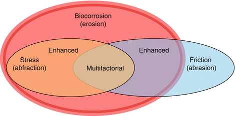
Fig. 14.3
Three conditions that are considered predisposing factors to NCCL formation are biocorrosion (erosion), abrasion, and occlusal stresses. The contribution to NCCL formation of both abrasion and occlusal stresses is enhanced in the presence of an acidic environment; whereas, when occlusal stresses and abrasion are combined outside of an acidic environment, there does not seem to be the same additive effect. And while biocorrosion or abrasion may at times seem to be the major single contributing factor in some cases of NCCLS, occlusal stresses when implicated are usually thought of as being in association with other factors rather than alone. The etiology of NCCLs is multifactorial [42]
14.3 Clinical Presentation of Noncarious Cervical Lesions
Noncarious cervical lesions may present on coronal tooth structure, a combination of coronal and radicular structure, or radicular structure only. Some authors have proposed that the morphological characteristics of noncarious cervical lesions are determined by their specific etiology [31].
Shallow defects on smooth surfaces coronal to the CEJ are considered to be a pathognomonic sign of biocorrosive tooth loss. U-shaped or disk-shaped broad shallow lesions with poorly defined margins and adjacent smooth enamel are consider to be erosive/biocorrosive lesions resulting from extrinsic acid sources such as those consumed through diet, medication, or recreational drug use. Extrinsic biocorrosive agents generally result in lesions on the facial surfaces of anterior teeth. On the other hand, intrinsic forms of biocorrosive lesions, caused by the reflux of gastric contents, are generally located on the lingual and incisal surfaces of maxillary anterior teeth.
Abrasive lesions generally exhibit sharply defined margins and a hard surface that may display traces of scratching.
Abfraction lesions are characterized as being irregular and may be wedge-shaped or saucer-shaped with sharp internal angles. However, the fact that the etiology of NCCLs is multifactorial makes differentiation between causative factors difficult. Acidity, abrasion, and tooth flexure each play a role to a greater or lesser degree from patient to patient and tooth to tooth, and so shape cannot be considered totally predictive of etiology.
14.4 Management of Noncarious Cervical Lesions
The decision on how or when to treat NCCLs varies widely among practitioners. In 2003, the Academy of Operative Dentistry published recommendations for the treatment of noncarious cervical lesions that should still serve as guidelines today [32]. The decision to restore these lesions depends upon the following factors:
1.
Inability to eliminate or greatly reduce the rate of lesion progression through elimination of etiologic factors.
2.
Lesion is esthetically unacceptable to the patient.
3.
Significant sensitivity of exposed dentin to cold liquids, food, and air that cannot be handled more conservatively.
4.
Depth of the lesion threatens the strength of the tooth and integrity of the coronal-radicular unit.
Once all of the contributing factors for noncarious cervical lesion formation have been identified, the first therapeutic measures to be considered should be directed at preventing new lesions and halting the progression of existing lesions. Some of the treatment options for NCCLs, depending upon their severity, include dentin desensitization, restoration, periodontal surgery, or some combination of the three.
14.4.1 Preventive Measures
If acid plays a significant role, sources should be identified and eliminated as much as possible. Dietary counseling should address such issues as the frequency of intake of acidic foods, as well as the buffering capacity of certain foods. When drinking acidic beverages, the use of a straw followed shortly afterward by rinsing the mouth or drinking milk or eating cheese should be encouraged. Patients with conditions such as gastric reflux or bulimia should be counseled to seek medical attention to attempt to elicit and treat the underlying causes. If elimination of the acid source is not feasible, the damage caused by the acid should be mitigated as soon as possible through buffering. The use of antacid lozenges, alkaline mouth rinses, or sugar free gum to stimulate salivary flow all help buffer acidity. Enhancing resistance against acid attack through the use of fluoride toothpaste, rinses, gels, and varnishes should also be considered as a part of a preventive treatment plan.
Identifying potential sources of abrasion to teeth should be investigated. Patients should be educated not only on proper oral hygiene techniques and types of preventive aids, but also on how and when to consider brushing. When teeth have had time to remineralize after an acid challenge, the potential for mineral loss can be minimized.
As discussed earlier, abfraction as a primary factor in causing NCCLs continues to be challenged [33]. Though laboratory studies have evaluated the role of occlusal forces in the formation of NCCLs, and have duplicated the cervical area on the tooth where stress seems concentrated [34], there is no consensus on treatment strategies. A review of the literature by Wood et al. [35] in 2008 found that there was no evidence to support occlusal adjustment as a being helpful in terms of slowing down lesion formation or improving the retention of restorations when placed to restore NCCLs [35]. A systematic review of 286 articles on this topic, by Senna et al. in 2012, found that only 28 articles met the criteria for review, and that much of the literature that was published were review articles that were merely reiterating literature that was both quantitatively and qualitatively weak in terms of types of study design. With that in mind, they reported that in the available literature, a causal relationship between NCCLs and occlusion had not yet been demonstrated by prospective studies, and that cross-sectional studies only very lightly support that contention [36]. They encouraged researchers to plan further studies to seek a causal relationship, taking into consideration the necessity of eliminating bias.
Therefore, trying to stop the initiation of new NCCLs or the progression of existing lesions by adjusting the occlusal forces on the tooth is not currently supported by the literature. Clinicians that try to do so must be cognizant that inappropriate occlusal adjustments may increase the risk for other occlusal problems and that they should limit their adjustments to altering inclined cuspal inclines, reducing heavy contacts, and removing interferences as these help in reducing laterally directed stresses that are, in theory at least, the most damaging. Though controversial and not evidence based, the use of occlusal splints is another conservative option to try and mitigate the damage from those forces [37].
14.4.2 Dentin Desensitization
Dentin hypersensitivity has been referred to as one of the most painful and least successfully treated chronic dental conditions of teeth [38]. Exposed dentin in the cervical area may become hypersensitive, whether it is the result of loss of significant tooth structure or not. Exposure to normal thermal, evaporative, tactile, osmotic, or chemical stimuli can all result in exaggerated painful responses for patients with dentin hypersensitivity. It is important to distinguish dentinal sensitivity pain, which is short in duration from pain of longer duration, which may be the result of pulpal inflammation. The most widely accepted explanation of dentinal sensitivity is the hydrodynamic theory [39]. The basis of this theory is that changes in direction of fluid movement within open dentinal tubules are perceived as pain by mechanoreceptors near the pulp. Under normal conditions, dentin is not exposed to the oral environment because it is covered either by cementum or enamel. In the case on NCCLs, that protective layer is removed and dentin becomes exposed. However, not all exposed dentin becomes sensitive. For pain to be experienced, the dentin tubules must be open at the surface. Studies have demonstrated that sensitive dentin contains as many as eight times as many open or patent tubules per unit area than nonsensitive dentin. Tubule diameters in sensitive teeth were also twice as wide in diameter [40].
Traditionally, the therapy for management of dentin hypersensitivity is primarily aimed at either occluding the dentinal tubules, creating coagulates inside the tubules to stop or minimize fluid movement within them, or by interfering with the transmission of pain signal at the synapse (typically with potassium nitrate). Diverse agents or formulations have shown various degrees of effectiveness in reducing the symptoms of dentin hypersensitivity, with some being applied professionally and others being applied as at-home treatments. A randomized clinical effectiveness study published in 2013 evaluated the comparative effectiveness of three treatments for hypersensitive NCCLs over 6 months. Potassium nitrate dentifrices, placement of a resin composite restoration, and placement of a sealant (DBA) were compared. The sealant and restoration proved equally effective in reducing hypersensitivity, while the dentifrice reduced hypersensitivity, but over time. The study recommended long-term studies of 3–5 years to determine the most effective treatment modality [41].
An in-depth discussion on dentin hypersensitivity can be found in Chap. 15 of this book.
14.4.3 Restoration of NCCLs
When desensitization of the tooth is not enough or there are esthetic or structural integrity issues that need to be considered for the tooth, a restoration may become necessary. Whether or not a restoration is truly necessary should be weighed heavily against the slowly progressing nature of the lesion and the high capacity of the patient’s self defense mechanisms for sclerotic dentin formation since restoration failure could lead to a cycle of rerestoration that would require restoration replacement numerous times throughout the patient’s life. This is especially important in light of the fact these restorations can be some of the least predictable or durable restorations that are placed (Fig. 14.4).
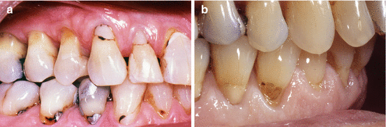
Fig. 14.4
NCCL restorations are prone to recurrent caries (a) and dislodgment (b)
The challenges presented by material weaknesses as well as the lesions’ cervical location and the inherent presence of variable bonding substrates, the restorative material’s elastic modulus, polymerization shrinkage, and lack of resistance to wear and erosion can all compromise clinical longevity. Additional contributing factors to failure include the fact that cervical lesions are not prepared to include any macromechanical retention. In addition, they often present a bonding surface that is mostly in dentin, and often involving sclerotic dentin in an area of the tooth that is difficult to isolate. Therefore, restoration failure is likely due to the combined effect of insufficient material properties, the continued presence of the etiological factors that caused the lesion initially, and the specific biological environment in the cervical area [42].
Stay updated, free dental videos. Join our Telegram channel
VIDEdental - Online dental courses


