Introduction
The aim of this article was to present a new method of analysis for the assessment of facial growth and morphology after surgical resection of the mandible in a growing patient.
Methods
This was a 2-year longitudinal study of facial growth in a child who had undergone segmental resection of the mandible with immediate reconstruction as a treatment for juvenile aggressive fibromatosis. Three-dimensional digital stereo-photogrammteric cameras were used for image acquisition at several follow-up intervals: immediate, 6 months, and 2 years postresection. After processing and superimposition, shell-to-shell deviation maps were used for the analysis of the facial growth pattern and its deviation from normal growth. The changes were seen as mean surface changes and color maps. An average constructed female face from a previous study was used as a reference for a normal growth pattern.
Results
The patient showed significant growth during this period. Positive changes took place around the nose, lateral brow area, and lower lip and chin, whereas negative changes were evident at the lower lips and cheeks area. An increase in the vertical dimension of the face at the chin region was also seen prominently.
Conclusions
Three-dimensional digital stereo-photogrammetry can be used as an objective, noninvasive method for quantifying and monitoring facial growth and its abnormalities.
Facial changes during growth have always been of interest to orthodontists and maxillofacial and plastic surgeons. In the past, clinicians have always relied on photographs for preoperative treatment planning in an attempt to restore the “normal” face. Facial photographs have also been used for postoperative assessment, medicolegal documentation, and direct communication with patients. The study and the evaluation of soft tissues of the face play integral roles in the analysis and description of variable patterns of craniofacial growth. Craniofacial anthropometry has been limited to direct measurements on subjects and has been shown to be reliable and inexpensive. However, this technique is time consuming and requires the patient’s compliance, which is difficult to achieve in children. Presently, several methods are available for capturing and quantifying craniofacial surface morphology, examples of which are 2-dimensional photogrammetry and more recently 3-dimensional (3D) methods. Three-dimensional methods overcome the limitations and measurement errors of 2-dimensional methods because 3D surfaces are represented. Soft-tissue 3D captures of the face can be accomplished with laser surface scanners or, more recently, with digital 3D stereo-photogrammetry, which has the advantages of showing color and texture and requiring less capturing time. It is therefore used more with children and developmentally impaired subjects (who are unable to maintain their posture for a period of time). Several 3D stereo-photogrammetric systems have been described in the literature. Among the most commonly described are the C3D imaging system (Glasgow, Scotland), Genex 3D camera system (Bethesda, Md), and the 3dMDface system (Atlanta, Ga). The reliability and validity of craniofacial anthropometric measurements with 3D photogrammetric images have been extensively tested and found to be reliable with a high precision and accuracy. Software tools are available that allow for the manipulation of images by the user to facilitate the identification of landmarks and the calculation of anthropometric measurements. The ability to capture 3D images has opened up new avenues for objectively analyzing craniofacial changes. The wide application of 3D imaging has been discussed in the literature; extensive studies have been done for the assessment of facial growth by calculating the displacement vectors of each landmark for determining both the effects of aging and sex. Other studies have compared soft-tissue changes, and lip and nasal morphology in patients who have undergone surgery for cleft lip and palate against a set of control subjects. Facial scars in children who were burned and the amount of soft tissue needed by using a soft-tissue expander to cover the defect have been determined with 3D facial photogrammetric imaging. Monitoring of craniofacial growth, presurgical and postsurgical soft-tissue assessment, and objective evaluations of soft-tissue changes and relapse in orthognathic surgery can be achieved now by using 3D stereo-photogrammetry.
Normal growth and posturing of the mandible are essential for the normal development and growth of the face. Disruption of mandibular continuity and mandibular defects can affect the coupling of the maxillomandibular growth pattern. Thus, considerable deformity and dysfunction can result from surgical segmental resection of the mandible in a growing child. Reconstruction of a pediatric mandible is considered challenging because of the unpredictable growth pattern of the reconstructed side and its effect on the overall growth of the face.
To our knowledge, no studies have used 3D digital stereo-photogrammetry to evaluate the effect of segmental mandibular resection on facial growth in a pediatric patient. We present a study with the 3dMDface system to report the facial growth and longitudinal changes in our patient 2 years after the surgical resection and the comparison with a normal growth pattern.
Material and methods
A 9-year-old white girl, who had undergone segmental resection of the left side of the mandible with preservation of the condylar head, is described in this report. The resection was made as a treatment for a mandibular lesion diagnosed as juvenile aggressive fibromatosis (JAF) as confirmed by the biopsy and the pathology report. JAF is a locally aggressive neoplasia with a high recurrence rate; it grows rapidly and can mimic cancer but does not metastasize. It affects infants and children and requires radical surgery. A complete literature review including all case reports of JAF between 1960 and 2003 was presented by Spere et al.
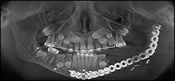
The patient came to the Oral and Maxillofacial Department at the Memorial Herman Hospital of Harris County in Houston, Texas. The lesion extended from the distal end of the left deciduous second molar along the ramus to the neck of the left condyle. Radical resection of the lesion with a safe margin of 2 to 3 cm by using a lower-lip split technique was performed by an author (J.W.) from the maxillofacial surgery team under general anesthesia with nasal intubation. Immediate reconstruction of the defect was made by using a 2.4-mm syntheses titanium reconstruction plate fixated with 6 screws placed anteriorly and 2 screws proximally in the head and neck of the left condyle for rigid fixation ( Fig 1 ). Orthodontic treatment was initiated for this patient to maintain the correct relationship of the dental arches and control the eruption and function of the remaining dentition. Several bone markers were placed at the time of operation for monitoring mandibular growth and its displacement vectors. Three-dimensional surface scans with the 3dMDface system were taken at several follow up times: immediately after resection (T1), 6 months after resection (T2), and 2 years after resection (T3) to monitor and evaluate the soft-tissue changes during dentofacial growth and the changes in the mandibular growth pattern after resection. The child was 11 years old at T3. Her body mass index was within the normal range.
The scans of 26 nonsyndromic girls from a previous longitudinal study, who had not received orthodontic treatment and had no craniofacial anomalies, were combined to produce an average face. The average face was created by aligning all faces in a group along a predefined axis of space by using the closest-point algorithm as previously described. The growth pattern, outlining the 3D shape of the patient, was compared with the growth of the average constructed face.
The digital stereo-photogrammetric 3dMDface system uses a noninvasive method for surface acquisition; it consists of precisely synchronized cameras positioned as a stereo pair in an optimum configuration. A random light projection is used to capture an image from the synchronized digital cameras at multiple angles to reconstruct a digital 3D image. The craniofacial image is visualized as a collection of points in 3D space resulting from the reconstructed craniofacial surface. One continuous point cloud is produced from the 2 stereo camera viewpoints; this eliminates data errors associated with merging or stitching data sets together. These points in the x, y, and z coordinate system are interrelated, and the distances among these points can be readily computed to produce the surface image. The system captures full facial images from ear to ear in less than 1.5 ms, producing color images with a resolution up to 40,000 points per square inch with minimal motion distortion because of the fast capturing time, making this system useful in children. The manufacturer’s stated accuracy is less than 0.2 mm, and the clinical accuracy quoted is 1.5% of the total observed variance. Images taken by the 3dMDface system were viewed on a computer by using the 3dMDpatient software platform.
The patient was seated on an adjustable stool and asked to look at an object located centrally between the 2 cameras; adjustments were made to achieve natural head posture. This position allowed the patient to be relaxed and the facial soft tissues to be in a state of least tension. This technique has been proven to be clinically reproducible. Each image took approximately 50 ms to be captured, recording the features of the surface of the face by means of triangulation and transferring them to the 3dMD modular system software on a computer workstation and converting them into a 3D image.
The images were imported and analyzed by using the Rapidform2006 (RF6) software. The data were further processed before analysis to obtain an image that preserved shape, surface, and volume by using customized macros built in the RF6. Once this was accomplished, a final facial shell was created for each scan. The absolute mean shell deviations, standard deviations of errors during shell-to-shell overlaps, maximum and minimum range maps, histogram plots, and color maps were generated by using the RF6.
All 3 scans at T1, T2, and T3 were superimposed to determine the changes taking place at the various times, with T1 as the reference template. This systematic process was done by manually aligning 5 points on the facial scans (2 points at the inner canthus of the eyes, 2 at the corners of the mouth, and 1 point on the nasal tip). This was followed by fine registration, when the software RF6 determined the best fit of the 2 scans. The closest-point algorithm, or best-fit method, was used and has been described previously.
A quantities analysis to determine the magnitude of changes between 2 shells was made by using shell-to-shell deviation maps after shell superimposition. The shell-to-shell deviation maps pooled all surface changes and produced a mean absolute value. Color histograms produced by the RF6 software showed the areas of change between the facial shells, with the added benefit of showing the directions of change (ie, positive and negative). These colored histograms showed surface changes that had increased or decreased between T1, T2, and T3. These changes were analyzed in millimeters and as percentages.
Stay updated, free dental videos. Join our Telegram channel
VIDEdental - Online dental courses


