Supernumerary teeth are an infrequent developmental anomaly that can appear in any area of the dental arch and can affect any dental organ. Multiple supernumerary teeth, or hyperdontia, is rare in people with no other associated diseases or syndromes. Conditions commonly associated with hyperdontia include cleft lip and palate, trichorhinophalangeal syndrome, cleidocranial dysplasia, and Gardner’s syndrome. A black girl, aged 11 years 8 months, came for consultation; radiographs showed 81 teeth: 18 deciduous, 32 permanent, and 31 supernumerary. The main concern initially was to determine whether she was syndromic, and she was referred to a geneticist. G banding analysis showed pericentric inversion of chromosome 9; the chromosome formula was 46, XX, inv (9) (p13q21). Orthodontic treatment for this patient will be a clinical challenge because of the great number of teeth to be extracted and the alterations in the shapes of the teeth. Treatment goals should be established by a multidisciplinary team, where oral surgeon, orthodontist, periodontist, and prosthodontist come together to solve a medical and dental puzzle, eliminating the pieces that do not fit and searching for new ones to obtain an occlusion that will give the patient physiologic conditions of normality and esthetic satisfaction.
Dental anomalies occur because of a variety of genetic and environmental factors. Combinations of dental anomalies are associated with specific syndromes. A few cases of multiple dental anomalies have been reported in patients with no generalized abnormalities.
The presence of supernumerary teeth, or hyperdontia, is an infrequent developmental alteration that appears in any area of the dental arches and can affect any dental organ. The etiology of supernumerary teeth is not completely understood. There are various theories for the different types of supernumerary teeth. One theory suggests that a supernumerary tooth is created as a result of a dichotomy of the tooth bud. Another theory, well supported in the literature, is the hyperactivity theory, which suggests that supernumeraries are formed as a result of local, independent, and conditioned hyperactivity of the dental lamina. Heredity might also play a role in this anomaly.
Occurrence can be single or multiple, unilateral or bilateral, erupted or impacted, and in 1 jaw or both jaws. Multiple supernumerary teeth are rare in people with no other associated diseases or syndromes. The conditions commonly associated with an increased prevalence of supernumerary teeth include cleft lip and palate, trichorhinophalangeal syndrome, cleidocranial dysplasia, and Gardner’s syndrome.
Although missing and supernumerary teeth are asymptomatic in most cases, they can lead to malocclusions, and esthetic, functional, and psychological problems. The presence of supernumerary teeth is associated with different alterations in neighboring teeth; the most common are overretained teeth or delayed eruption, ectopic eruption, dental malposition, occlusal problems, diastemas, and rotated neighboring teeth, among a series of associated pathologies. An early diagnosis prevents or reduces the risk of complications and, when combined with early removal, has a better prognosis. Removal of a supernumerary tooth preventing permanent tooth eruption usually slloed the eruption of the tooth, if adequate space is available in the arch to accommodate it.
This article presents an unusual case of multiple hyperodontia in a girl aged 11 years 8 months with 31 supernumerary teeth.
Case report
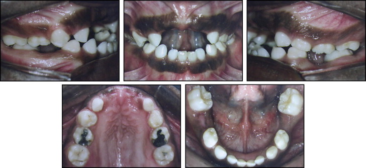
A black girl, aged 11 years 8 months, came for consultation for removal of root fragments of the maxillary deciduous incisors. During the clinical examination, it was noted that her only permanent teeth were the 4 first molars and the maxillary right first premolar. The following deciduous teeth were present in the oral cavity: 53, 55, 63 through 65, 71 through 74, and 81 through 84 ( Fig 1 ).
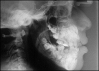
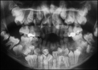
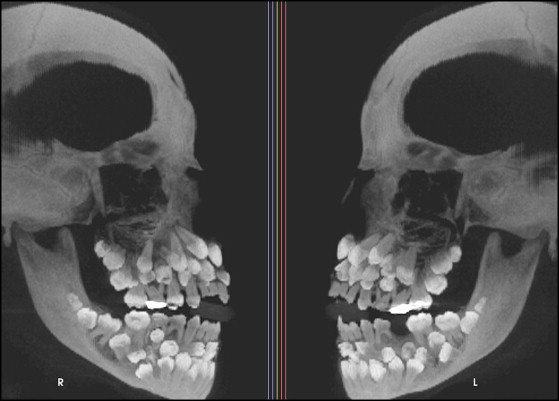
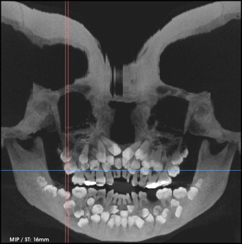
Panoramic and lateral cephalometric radiographs were requested, and they showed a significant clinical finding: multiple supernumerary teeth ( Figs 2 and 3 ). Cone-beam computed tomography (CBCT) was ordered to evaluate 3-dimensionally the number and the correct positions of the supernumerary teeth. The CBCT images showed a total of 81 teeth: 18 deciduous, 32 permanent, and 31 supernumerary ( Figs 4 and 5 ). The scans showed alterations of the forms of the teeth; this made it difficult to differentiate between the supernumerary and the other teeth.
Cervical and thorax computed tomography images showed normal osseous structures, no expansive lesions, and intact muscular-adipose planes. During the medical history evaluation, it was found that the patient’s mother was thalassemic; however, no alteration in her number of teeth was found.
Discussion
Because multiple supernumerary teeth are associated with diseases or syndromes, a thorough study should be conducted to establish a differential diagnosis in such patients. The presence of 31 supernumerary teeth caused suspicion. No case of a genetic syndrome in the family related to hyperdontia was reported in the patient’s medical history. The fact that her mother was thalassemic did not seem to be relevant, because supernumerary teeth are not a characteristic of this type of anemia. Tooth crown sizes (mesiodistal and buccolingual diameters) of thalassemic male and female patients were significantly smaller than those of unaffected control subjects. Beta-thalassemia major is associated with greater dental caries experience.
The conditions commonly associated with an increased prevalence of supernumerary teeth are cleft lip and palate, trichorhinophalangeal syndrome, cleidocranial dysplasia, and Gardner’s syndrome. During the clinical evaluation, it was noted that the patient did not have cleft lip and palate.
Cleidocranial dysostosis is a rare congenital disorder of bone with an autosomal dominant hereditary mode of inheritance. This condition is characterized by clavicular aplasia or deficient formation of the clavicles, delayed and imperfect ossification of the cranium, moderately short stature, and a variety of other skeletal abnormalities. The oral manifestations are delayed exfoliation of deciduous teeth, delayed or failed eruption of the permanent dentition, and multiple supernumerary teeth. The most characteristic and pathognomonic feature of this disorder is hypoplasia or aplasia of the clavicles, which results in hypermobility of the shoulders, allowing the patient to approximate the shoulders in the midline.
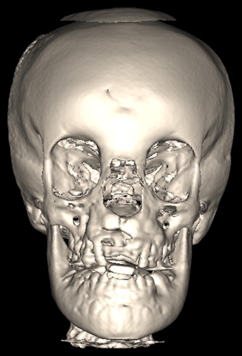
Of the characteristics commonly found in patients with cleidocranial dysostosis, only oral manifestations were present. Clavicular disorders could not be verified. The CBCT images showed hyperostosis in the region of the frontal bone ( Figs 6 and 7 ).