Introduction
Our objective was to study mandibular widening in untreated subjects with hemifacial microsomia.
Methods
From the 3-dimensional files at the Department of Plastic and Reconstructive Surgery, Skane University Hospital in Malmö, Sweden, data of 11 subjects (3 girls, 8 boys) with hemifacial microsomia were retrieved. Their age range was 9 years 2 months to 13 years 2 months at the first examination. The mean observation period was 5 years 2 months. Each subject was studied by roentgen stereophotogrammetry with the aid of metallic implants.
Results
A significant widening of the mandible was found, with a mean total change of 0.31 mm (range, 0.08-0.79 mm) and a mean annual change of 0.07 mm (range, 0.03-0.12 mm). In 2 subjects, narrowing of the mandible was found: −0.16 and − 0.23 mm.
Conclusions
The mandible got wider during adolescence in 9 of our subjects with hemifacial microsomia but to a lesser extent than has been reported in subjects without hemifacial microsomia and from ordinary orthodontic clinics. Sex difference was not addressed. It was suggested that chewing (forces and patterns) was responsible for the mandibular widening in our subjects; this is in line with previous research.
The mandibular symphysis fuses before the end of the first year of life. The mandibular corpus-ramus grows backward by bone apposition at the posterior border and resorption at the anterior border of the ramus. These 2 concepts are well established in the literature about mandibular growth. Moreover, mandibular growth reaches its peak during puberty.
Baumrind and Korn and Iseri and Solow reported mandibular widening with age, but on different grounds. Baumrind and Korn suggested that the symphysis acted as a vertical hinge axis, whereas Iseri and Solow suggested that chewing forces caused the widening. Both studies used conventional profile radiographic images (combining profile and posteroanterior roentgenograms) and metallic implants. The cephalostat was used for calibration. In both studies, they used subjects (girls and boys) from ordinary orthodontic clinics. Sex differences were not addressed in either study. Our long-standing experience with roentgen stereometric analysis prompted us to examine our data on subjects with hemifacial microsomia (3 girls, 8 boys) to determine whether they showed mandibular widening as has been reported in subjects without hemifacial microsomia. Roentgen stereometric analysis is at present, and to our knowledge, the only 3-dimensional method with which real linear and angular measurements based on implants exclusively can be recorded with high precision and accuracy. Roentgen stereometric analysis does not use skeletal reference points and is thus independent of sutural growth or remodeling (resorption or apposition) due to function. We have used this method in several studies from 1975 through 2004.
Material and methods
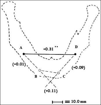
From the 3-dimensional craniofacial database at the Department of Plastic and Reconstructive Surgery, Skane University Hospital in Malmö, Sweden, 11 untreated subjects (3 girls, 8 boys) with hemifacial microsomia examined before and after puberty were selected. Four mandibular implants were required, forming a quadrangle, ABCD ( Fig ). The subjects’ age range was 9 years 2 months to 13 years 2 months at the first examination. The mean observation period was 5 years 2 months. These subjects had been included in earlier studies.

Since 1975, facial skeletal growth in children with craniofacial anomalies has been examined by roentgen stereometric analysis at the Department of Plastic and Reconstructive Surgery and the Centre of Craniofacial Anomalies, Skåne University Hospital, Malmö, Sweden. This was approved by the committee of ethics of the Faculty of Medicine, University of Lund (June 19, 1975), with the consent of parents and patients. Roentgen stereometric analysis with the aid of metallic implants was developed by Selvik for the study of the kinematics of the skeletal system: ie, facial bones. For calibration, a cage with reference indicators is used. At each examination, the patient’s head is placed in the cage, and 2 x-ray units at approximately right angles are exposed simultaneously. The examinations were performed irregularly because of clinical requirements. The 3-dimensional data are obtained by digitizing both the implants of the mandible and those of the calibration cage (accuracy, 0.01 mm 4 ); with the aid of a television display, the coordinates are fed into the computer.
Balls (0.5 mm) or pins (1.5 × 0.5 mm) are routinely inserted mesially to the canine and at the level of the mandibular first molar bilaterally ( Fig ) and used for the study of mandibular displacements. The 4 implants formed the quadrangle ABCD.
The stability of implants is routinely checked at each examination by calculation of angles and distances of the quadrangle ABCD. The limits for mean errors of the triangles ABC and BCD ( Fig ) should be less than 0.2 mm and 0.4°, respectively; these are the limits for implant stability from Selvik and Rune et al. If these limits are passed, the implants are unstable, and there are 2 options: to insert an additional implant or to leave it out in the subsequent analysis if there are still 3 implants left, to determine the position of the bone in space. If an additional implant is inserted, a new series of examinations begins. In this study, the A to D distance corresponded with the “body implant” of Baumrind and Korn and the “ipl-ipr” of Iseri and Solow. The 2 implant lines, A to B and C to D, formed the interimplant line angle: “hinge-axis angle” (by the subtraction of the supplementary angles ABC and BCD from 180°).
Statistical analysis
Three-dimensional data (linear and angular) of the 11 subjects, retrieved from our craniofacial database, represented the growth period. For each subject, the total and annual changes of mandibular width (A-D) and the total changes of the ABC and BCD angles were retrieved. The Student t test for paired observations was used in the statistical analyses.
Results
The total and annual changes in mandibular width are presented in Table I . The total width (A-D) increased in 9 subjects (range, 0.08-0.79 mm). The mean total linear change of 0.31 mm (SD, 0.024) was significant at the 5% level. The mean annual linear change was 0.07 mm (range, 0.03-0.12 mm). In 2 subjects (412 male [M], 433 female [F]), the total width (A-D) decreased by −0.23 and −0.16 mm, respectively, with an annual linear change of 0.04 mm in both. The mean total linear changes of the remaining 3 sides were A-B, +0.01; B-C, +0.11; and C-D, +0.09 mm. These changes were not significant ( Fig ).
| Subject | Total/annual (mm) | Observation period (y, mo) |
|---|---|---|
| 412M | −0.23/0.04 | 11, 9-16, 6 |
| 433F | −0.16/0.04 | 13, 2-17, 10 |
| 440F | +0.25/0.08 | 13, 0-16, 3 |
| 441M | +0.41/0.10 | 11, 2-15, 2 |
| 444M | +0.47/0.08 | 12, 1-18, 3 |
| 447M | +0.79/0.12 | 10, 3-16, 11 |
| 488M | +0.62/0.10 | 9, 2-15, 9 |
| 500M | +0.30/0.10 | 9, 10-13, 0 |
| 505M | +0.08/0.03 | 12, 4-15, 8 |
| 508F | +0.22/0.03 | 10, 1-16, 11 |
| 511M | +0.09/0.04 | 10, 5-13, 0 |
| Mean | +0.31/0.07 | 52 |
| SD | 0.024 | Range, 9, 2-18, 3 |
| SEM difference | 0.083 | |
| Range | −0.23 to +0.79 | t value, 3.8 ∗ |
Angular changes of ABC, BCD, and the hinge axis are presented in Table II . Only 1 subject (488M) showed increases of both the ABC and BCD angles, indicating widening of the mandible (3.4° and 1.3°). Six subjects showed significant changes in opposite directions (412M, 440F, 441M, 444M, 500M, and 508F), and 4 subjects showed just minor changes in at least 1 parameter. The hinge axis increased in 4 subjects (412M, 441M, 488M, and 500M) and decreased in 2 (433F, 447M). In the remaining 5 subjects, the changes were less than 0.4 mm.


