Introduction
Most of the published literature on the management of overbite with the Invisalign appliance (Align Technology, Santa Clara, Calif) consists of case reports and case series.
Methods
In this retrospective study of 120 patients, we sought to assess the nature of overbite changes with the Invisalign appliance. Records were collected from 3 practitioners, all experienced with the Invisalign technique. The patients were consecutively treated adults (>18 years old) who underwent orthodontic treatment only with the Invisalign appliance. Patients with major transverse or anteroposterior changes or extraction treatment plans were excluded. The study sample included 68 patients with normal overbites, 40 with deepbites, and 12 with open bites. Their median age was 33 years, and 70% of the patients were women.
Results
Cephalometric analyses indicated that the deepbite patients had a median overbite opening of 1.5 mm, whereas the open bite patients had a median deepening of 1.5 mm. The median change for the normal overbite patients was 0.3 mm. Changes in incisor position were responsible for most of the improvements in the deepbite and open bite groups. Minimal changes in molar vertical position and mandibular plane angle were noted.
Conclusions
The Invisalign appliance appears to manage the vertical dimension relatively well, and the primary mechanism is via incisor movements.
Highlights
- •
The Invisalign appliance is relatively successful in managing overbite.
- •
Our results did not show that posterior teeth intrude significantly with the Invisalign appliance.
- •
The Invisalign appliance improved deepbites primarily by proclination of the mandibular incisors.
- •
The Invisalign appliance corrected mild to moderate anterior open bites mainly through incisor extrusion.
The Invisalign appliance (Align Technology, Santa Clara, Calif) consists of a series of computer-designed clear plastic shells that fit closely over the teeth and incrementally move the teeth to their correct position. Orthodontic treatment with the Invisalign appliance may be more esthetically appealing to some patients when compared with conventional fixed appliances; this partly explains the increasing demand for this treatment method.
The Invisalign technique was initially proposed to treat mild orthodontic cases. Nonetheless, there are reports of complex orthodontic cases treated with the Invisalign appliance in the literature. For example, a recent study demonstrated the successful closure of a 4-mm anterior open bite by extrusion of the anterior teeth using a series of 35 Invisalign aligners.
Soon after the introduction of the Invisalign system in the late 1990s, practitioners noticed that the appliance commonly induced deepening of the overbite. It was suggested that aligners covering all posterior teeth could function as a bite-block, thereby intruding the posterior teeth. This would result in a reduction of the posterior vertical dimension and consequent deepening of the overbite.
The Invisalign system has evolved over the last 16 years, and various strategies have been developed to better manage the vertical dimension. For example, an early strategy to prevent bite deepening was the removal of occlusal coverage on the second molars. Align Technology recently developed new treatment options including specially designed attachments and virtual bite ramps. Attachments are composite buttons attached to the labial surfaces of the teeth, and they come in various shapes to assist with tooth movement. Specifically, these attachments increase retention, transmit desirable force to the teeth, and support auxiliary functions such as placement of elastics. Virtual bite ramps function similar to bite plates or bite turbos. These bite ramps, incorporated into the maxillary aligner, contact the mandibular incisors to disocclude the posterior teeth when patients bring their teeth together.
Despite these advancements in the Invisalign appliance, evidence supporting the effectiveness of these treatment modalities is limited to case reports and case series. Studies with larger samples and better designs are required to understand the mechanism by which the Invisalign appliance manages the vertical dimension. In this retrospective study, we sought to investigate the vertical dimension changes in patients with various pretreatment overbite relationships treated only with the Invisalign appliance. Moreover, we aimed to identify the dental and skeletal changes associated with bite closing or opening. See Supplemental Materials for a short video presentation about this study.
Material and methods
This study was approved by the institutional review board of the University of Washington.
The study sample consisted of adult patients consecutively treated with the Invisalign appliance in 3 private orthodontic offices. Two practices were located in the greater Seattle area, Wash and one Vancouver, British Columbia. A total of 313 patient records were screened; records of 193 patients (62%) were excluded. The most common reason for exclusion was lack of final lateral cephalometric radiographs. The second most common reason was that the posterior teeth were out of occlusion when the radiograph was taken.
The study sample was stratified into groups of normal overbite, deepbite, and open bite based on the pretreatment overbite measured on cephalometric radiographs. Normal overbite was defined as pretreatment overbite ranging from 0 mm to less than 4 mm. Patients with 4 mm or greater pretreatment overbite were classified in the deepbite group. The open bite group included patients with negative pretreatment overbite.
Inclusion criteria were (1) the patient was 18 years or older at the beginning of treatment, (2) the treatment was completed between January 1, 2010, and January 1, 2014, (3) 11 to 40 aligners were used for each arch, (4) a maximum of 3 revision sets of aligners was used, (5) the treatment plan was nonextraction, (6) the molar anteroposterior occlusal relationship was not changed (eg, no Class II to Class I occlusion change), (7) posterior-transverse relationships were not changed significantly (eg, no correction of posterior crossbite), (8) fixed appliances were not used, and (9) the patient had good-quality pretreatment and posttreatment cephalometric radiographs. Two investigators (R.K. and W.L.) screened consecutively treated patients at each orthodontic office. Each subject eligible for the study was then assigned an anonymous identification number, and the records were deidentified with these numbers.
The collected records included (1) pretreatment and posttreatment lateral cephalometric radiographs, (2) the Invisalign Treatment Overview form with information regarding number and location of attachments as well as potential interproximal reduction plans, (3) patient’s age at the start of the treatment, (4) patient’s sex, and (5) questionnaires filled out by the clinicians regarding their treatment strategies.
Deidentified lateral cephalometric radiographs were imported into software (Dolphin Imaging, Chatsworth, Calif) to perform cephalometric analyses. Seventeen landmarks were marked on the initial and final lateral cephalometric radiographs. We opted to mark the landmarks for the pretreatment and posttreatment radiographs of each patient sequentially to reduce potential landmark identification error. The software then calculated the linear and angular measurements, which were used in our statistical analyses.
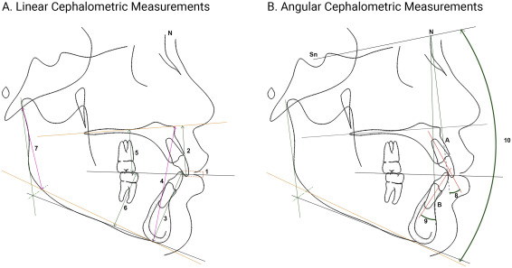
To assess the changes during treatment, 9 linear and 3 angular measurements were measured ( Fig 1 ). Palatal, occlusal, and mandibular planes were used as the reference lines. The palatal plane was defined as a straight line passing through the anterior and posterior nasal spine points. The occlusal plane was defined as a straight line drawn through the bisection of the mesiobuccal cusp tips of the first molars and the bisection of the incisal edge of the most anterior central incisors. The mandibular plane was defined as a straight line connecting menton to constructed gonion.

To assess the changes in the anterior vertical dimensions, we made these measurements. The linear measurements were overbite, defined as the shortest vertical distance between the tip of the maxillary incisor and the tip of the mandibular incisor perpendicular to the occlusal plane, and the vertical position of the incisors, defined as the shortest distance between the maxillary and mandibular incisors to the palatal and mandibular planes, respectively (reference lines). Additionally, the anterior facial linear height was measured: defined as the shortest distance between defined as the shortest distance between anterior nasal spine and menton was measured.
The angular measurements were the angle between the maxillary incisor’s long axis and the nasion-A point line and the angle between the mandibular incisor’s long axis and the nasion-B point line.
To assess the changes in the posterior vertical dimension, several linear and angular measurements were measured. The vertical dimension changes of the maxillary and mandibular molars were determined using linear measurements. The shortest distances between the palatal plane and the maxillary first and second molars’ mesiobuccal cusp tips were measured. Similarly, the shortest distances between the mandibular first and second molars’ mesiobuccal cusp tips to the mandibular plane were measured. The posterior facial height was measured as the shortest distance between constructed gonion and articulare. The angle between the mandibular plane and the sella-nasion reference line was measured.
Approximately 2 weeks after the initial measurements we, 10 cephalometric radiographs were randomly selected by the R statistical package (version 2.11.1; RStudio, Boston, Mass) through RStudio (version 0.99.491) for the measurement error analysis. Landmarks on these lateral cephalometric radiographs were reidentified, and the measurements were recalculated using the Dolphin Imaging software. The measurement error was reported as the mean difference between the initial and retraced cephalometric values.
Statistical analysis
Statistical analyses were conducted in 2 phases. Initially, descriptive analyses were performed to examine the cephalometric measurements of the pretreatment and posttreatment radiographs in all 3 groups. Detailed descriptive analyses are presented in the Appendix .
To examine the difference between cephalometric measurements before and after treatment, we used the nonparametric Wilcoxon signed rank test at the P = 0.05 level of significance. We opted to use this analysis because the majority of our variables were not normally distributed. Additionally, Kruskal-Wallis analysis at the P = 0.05 level of significance was used to investigate overbite changes in the 3 groups.
The statistical analyses were conducted using the R statistical package (RStudio).
Stay updated, free dental videos. Join our Telegram channel

VIDEdental - Online dental courses


