Bite wounds are especially prone to infectious complications, both local and systemic. In bite wounds to the face, such complications can create more difficulties than the initial tissue damage itself for the task of restoring an esthetic appearance. Management should aim to neutralize this potential for infection and provide an infection-free environment for wound healing. Wound cleansing followed by primary closure is the treatment of choice, and the use of prophylactic antibiotics may further decrease the risk of infection. Delay in presentation beyond 24 hours is not necessarily a contraindication to immediate repair, but excessive crushing of the tissues or extensive edema usually dictates a more conservative approach, such as delayed closure.
Bite wounds have always been considered complex injuries contaminated with a unique polymicrobial inoculum. Because wounds of the extremities constitute the majority of bite cases, most relevant studies have focused on the wound infection rate in these areas. However, a substantial subset of dog, cat, and human bites, each in the order of 15%, are located on the face, where fear of potential disfigurement is an overriding concern and the associated psychological consequences can be devastating.
Although a wide range of mammals have been implicated in facial bite injuries, the majority of these injuries are inflicted by dogs. It is estimated that there are 44,000 facial injuries from dog bites affecting children each year in the United States. Not surprisingly, facial injuries predominate in those dog-bite casualties requiring hospitalization.
For half a century, oral and maxillofacial surgeons have remained in the forefront of the surgical treatment of these injuries, with expertise in the pathogenic oral flora, due to their dental background. Nevertheless, certain aspects of therapy remain amenable to personal opinions and clinical impressions. The aim of this article is to discuss these issues in the general context of bite-wound management ( Box 1 ), including the role of prophylactic antibiotics and the possible limitations of the general axiom of primary closure.
- •
Selection of solution for wound irrigation
- •
Irrigation of puncture wounds
- •
Role of antibiotic prophylaxis
- •
Selection of antimicrobial agent(s)
- •
Cutoff time for primary closure
Wound characteristics
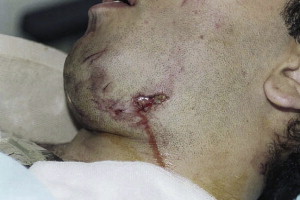
Animal bites can result in three main types of soft tissue trauma, namely punctures, lacerations, and avulsions, with or without an actual tissue defect. The typical dog bite results in a combination of torn tissues and adjacent punctures, the so-called “hole-and-tear” effect ( Fig. 1 ). Some degree of crush injury is also present in most bite wounds, including those from humans, due to the dynamics of the bite. Dog bites of the face are located mostly on the lips, nose, or cheeks. Human bites notably tend to involve the ear, although the lower lip is also prominently involved.
Bite wounds inflicted to the head and neck region by large animals can present in a more serious fashion. Large dog attacks can result in life-threatening or even fatal injuries because of airway compromise, exsanguination, or craniocerebral trauma. Furthermore, dog bites can impart enough energy to the facial skeleton to cause structural damage, especially in children.
Overview of microbiology
The importance of the indigenous oral bacteria in bite-wound infections is substantiated by the high isolation rates (>50% of cases) of Pasteurella spp from dog and cat bites, and viridans streptococci, especially Streptococcus anginosus , from human bites. There are also corresponding figures for oral anaerobes, including Fusobacterium nucleatum , Bacteroides , Prevotella , and Porphyromonas spp. It should be appreciated, however, that almost any oral organism can become a potential pathogen under the right circumstances.
Consistent with the heterogeneity observed between feline and canine oropharyngeal Pasteurella strains, P canis biotype 1 is the predominant isolate from dog bites, whereas P multocida subspecies multocida and septica have been isolated much more frequently from cat bites. Streptococci and staphylococci are the next most common aerobic isolates. Potentially invasive aerobic organisms isolated from domestic animal bites include Bergeyella ( Weeksella ) zoohelcum and Capnocytophaga canimorsus , the latter associated with fulminant systemic infections in immunocompromised hosts, usually after a dog bite.
Staphylococci are also commonly isolated from human bites. Eikenella corrodens , a normal inhabitant of the human oral cavity, appears to have a unique association with human bites, having been recovered from about 30% of cases. Other fastidious gram-negative organisms, such as Haemophilus spp and enteric gram-negative rods, have been found less frequently. Oral as well as environmental fungi may also contaminate bite wounds. Candida spp have been isolated from 8% of infected human bites, but their pathogenic role remains unclear.
Bites can also impart systemic bacterial and viral infections, including classic zoonoses. Human bites can be the source of the hepatitis B and C virus, and possibly HIV transmission, as well as syphilis. Rabies remains the most dreaded of all animal bite-wound infections, which should be especially considered when bites from bats, raccoons, or foxes are encountered.
Risk factors for wound infection
Facial bite wounds generally display low infection rates, commonly attributed to the rich blood supply of the area. Dog bites on the face are usually considered to be at moderate risk for infection when compared with other types of mammalian bites, especially those inflicted by cats, which harbor the more toxic P multocida organisms. Furthermore, dog-bite wounds seen within 3 hours of injury rarely contain more than 10 5 bacteria per gram of tissue, while human bites usually exceed this critical level because of higher bacterial counts in saliva.
Significant delays–beyond 6 to 12 hours–in seeking medical attention increase the likelihood of infection. Victims of bites to the face are more likely to present in time for prompt wound care than do other bite victims, because of concern about possible scarring. However, long delays may be encountered with facial bites, due to alcohol intoxication of the victim or transport from remote areas. Furthermore, prolonged exposure of the wound to bacterial contamination can affect the therapeutic efficacy of antibiotics. Unfortunately, no study has controlled for the time from wounding to antibiotic treatment.
Puncture wounds, typically inflicted by the slender feline teeth, are associated with high infection rates because they involve deep inoculation of pathogens. Crush injuries, on the other hand, can precipitate infection with significantly lower bacterial counts because of the resultant tissue ischemia. However, due to the inevitable cartilage exposure, avulsion injuries of the ear or nose inflicted by humans have the highest incidence of infection following facial bite wounds, according to reports.
Clinical evaluation
With extensive head or neck injury, life-preserving emergency procedures take precedence; cervical immobilization should also be considered. Otherwise, there is time to obtain the necessary information about the incident as well as about the general condition of the patient.
When there is a possibility of involvement of underlying specialized structures, early diagnosis is essential. Eyelid lacerations require careful evaluation to rule out penetrating injury to the globe or interruption of the lacrimal drainage system. Radiographic examination of the adjacent facial or cranial bones is indicated when a fracture is suspected. A proposed classification of facial bite wounds, based on extent, appears in Table 1 .
| Type | Clinical Findings |
|---|---|
| I | Superficial injury without muscle involvement |
| IIA | Deep injury with muscle involvement |
| IIB | Full-thickness injury of the cheek or lip with oral mucosal involvement (through-and-through wound) |
| IIIA | Deep injury with tissue defect (complete avulsion) |
| IIIB | Deep avulsive injury exposing nasal or auricular cartilage |
| IVA | Deep injury with severed facial nerve and/or parotid duct |
| IVB | Deep injury with concomitant bone fracture |
The wound should be assessed for signs of infection, including redness, swelling, or discharge. These signs tend to be more obvious with older wounds than with fresh ones. Fever is generally unlikely. P multocida organisms are associated with a rapid onset of infection, whereas when the latency period is more than 24 hours, staphylococci, streptococci, or anaerobes are more likely etiologic agents. Cultures are most useful in case initial antibiotic therapy fails.
Bite wounds are considered tetanus-prone, so appropriate immunization should be administered if the patient has had fewer than three doses of tetanus toxoid or more than 5 years have passed since the last dose. Rabies prophylaxis should be based on the local prevalence of the disease, the biting species, and the circumstances surrounding the incident.
Superficial bite wounds can be treated in the outpatient setting, whereas patients with more serious injuries (types III and IV) should be hospitalized and treated in the operating room. For children whose wounds require surgical care, hospitalization should be considered because they may be uncooperative under local anesthesia. Signs of systemic toxicity, rapidly advancing cellulitis, or infection despite oral antibiotic therapy constitute other indications for hospitalization. Most adults with uncomplicated bite wounds (type II) can be discharged after wound repair with instructions for follow-up.
Local wound care
As with any laceration, the mainstays of wound care are irrigation and removal of any necrotic tissue. However, common practices, such as cleansing with soap or scrubbing, are best reserved for high-risk wounds. Irrigation is essential in preventing infection because it removes debris and microorganisms; wounds difficult to irrigate thoroughly, such as punctures, are twice as likely to become infected. Manual irrigation with a 19-gauge catheter on a 30- to 60-mL syringe delivers a pressure range between 5 and 8 psi, considered optimal for appropriate decontamination. Continuous irrigation seems to be just as effective as pulsatile lavage. However, sustained high-pressure irrigation should be avoided in areas containing loose areolar tissue, such as the eyelids or children’s cheeks, because such irrigation may cause tissue disruption and excessive edema. In general, 250 to 500 mL of solution provides an adequate cleansing effect for most facial bite wounds. Although irrigation of puncture wounds remains controversial because of the inherent difficulties in proper drainage, most investigators also use pressure irrigation for these wounds, taking care to allow escape of the fluid ( Box 2 ). Incising the puncture to promote irrigation is not recommended, however, as it causes unnecessary scarring.
- 1.
Skin preparation; anesthesia
- 2.
Pressure irrigation; irrigation of puncture wounds
- 3.
Resection of skin tags
- 4.
Removal of visible foreign particles
- 5.
Suturing (exceptions listed below)
- 6.
Consideration of tetanus prophylaxis
- 7.
Follow-up within 24 to 48 hours
-
Also recommended:
-
Normal saline irrigation (1% povidone-iodine should be reserved for grossly contaminated wounds)
-
Antibiotic prophylaxis
-
Culture of problematic wounds (failure to respond to initial antibiotic therapy or presence of serious infection)
-
Not recommended:
-
Routine debridement (if attempted, it should not exceed 1 mm of tissue)
-
Suturing in the presence of overt infection, gross edema, foreign bodies, or visible contamination (consider delayed closure)
-
Culture of fresh uninfected wounds, because it depicts the polymicrobial flora of the wound rather than the causative organisms of any subsequent infection
Normal saline is the fluid of choice for irrigation, according to many experts. A 1% povidone-iodine solution also has been recommended for irrigation of bite wounds because this solution provides an optimal therapeutic balance between bactericidal capacity and tissue toxicity associated with iodine-containing formulations. However, when used under pressure for wound decontamination, saline has compared favorably with 1% povidone-iodine solution and other less commonly used alternatives. Moreover, even if povidone-iodine or another antiseptic solution is used as an irrigant, copious rinsing with normal saline should follow to minimize the risk of cytotoxicity.
Surgical debridement is a common clinical practice in bite-wound management because it significantly decreases the likelihood of infection. However, debridement of facial wounds should be kept to a minimum so as to avoid sacrifice of tissue that has a good chance to survive, particularly in landmark areas such as the vermilion border of the lips, the nasolabial fold, and the eyebrows ( Box 2 ).
Surgical treatment
Primary wound closure is the treatment of choice for all uninfected facial bite lacerations seen within 24 hours, as well as for many avulsion injuries, because this obtains the most favorable esthetic result. Subcutaneous sutures should be used sparingly, however, because they can act as foreign bodies and precipitate infection. By contrast, deep puncture wounds should be left open, particularly when inflicted by cats.
In the study of Maimaris and Quinton, 1 of 27 sutured wounds in the face became infected compared with none of the 14 wounds left open, a difference considered both insignificant and acceptable in view of the better cosmetic result achieved with suturing. Several other studies have confirmed the low risk associated with suturing of facial bite wounds, although in some studies increased infection rates were found both with dog bites and human bites.
For uncomplicated bite wounds presenting beyond the “golden 24-hour period,” primary closure is controversial. In these cases, delayed closure is a time-honored practice. This implies a waiting period of 4 to 5 days before definitive wound closure, during which time the wound is kept open, usually with moist gauze dressings providing drainage, while edema is allowed to subside. Antibiotics can be administered to further diminish the risk of infection.
Other surgeons, however, prefer to proceed with primary repair of late-presenting wounds to achieve a less noticeable scar, although this approach may increase the risk for infection. This approach has been substantiated by studies suggesting that primary closure of facial human bites can be undertaken with an acceptable risk within 48 hours and even as late as the fourth day after the incident. However, these studies included mainly low-risk wounds (ie, avulsion type rather than punctures or crush injuries), most of them located on the lips, which are very resistant to the development of infection.
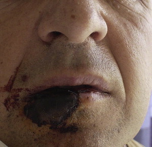
Avulsion bite wounds can pose reconstructive challenges if direct closure is not possible. Attempts to reattach avulsed parts are usually doomed to fail ( Fig. 2 ). In these cases, local skin flaps or composite grafts should be considered, depending on the area involved. Microsurgical replantation has become the standard operation in some centers, yet it remains technically demanding. Recently, an extensive soft tissue defect of the face due to a severe dog bite was reconstructed with partial face transplantation.
The presence of overt infection normally precludes suturing the wound. Options include secondary healing with subsequent revision surgery, delayed closure ( Box 2 ), or primary closure with insertion of a drain. Successful immediate primary closure has been reported after debridement with proteolytic agents.
Antibiotic treatment
Antibiotic administration for bite wounds can be either prophylactic or therapeutic. In the presence of established infection or any underlying predisposing condition, antibiotic therapy is indicated. However, it remains unclear whether otherwise healthy patients with fresh clinically uninfected wounds benefit from prophylactic antibiotic administration. Even in these cases, however, antibiotic therapy may actually be therapeutic if enough time has elapsed for bacterial proliferation to reach a level that can result in the development of infection.
On the basis of figures from a meta-analysis of prophylactic antibiotics for dog-bite wounds, Callaham calculated that as many as 26 patients must be treated with oral antibiotics to prevent 1 infection. Consistently, infection rates in the order of 4% have been reported with primary repair of facial dog-bite wounds without the use of antibiotics. On the other hand, with two notable exceptions, equally good results have been obtained when antibiotics were administrated. Obviously, little evidence supports the value of prophylactic antibiotics in the treatment of dog-bite wounds, although the type of wound, the particular location, and any additional contamination may necessitate antibiotic coverage.
Consensus exists regarding antibiotic prophylaxis for cat-bite wounds because of their high-risk character. Patients with human bites are also serious candidates for antibiotic prophylaxis. Limited evidence suggests that antibiotics for human bites of the face may result in infection rates as low as 2.5%. Furthermore, in a recent study, mainly dealing with high-risk avulsion injuries of the ear, failure to receive at least 48 hours of prophylactic intravenous antibiotics was associated with an increased infection risk following primary closure.
In view of the incomplete debridement permitted on the face, most investigators favor antibiotic prophylaxis for facial bite wounds because even low infection rates can seriously compromise cosmetic outcome, especially in children. Furthermore, it has been suggested that primary closure may also increase the risk of infection, thus further justifying prophylactic antibiotics in such cases. Because the indications for antibiotics do not correlate well with the severity of injury, prophylaxis is generally recommended for all bites penetrating the skin.
For most terrestrial mammal bites, the choice of antibiotics is based on experience with dog, cat, and human bites. Furthermore, because E corrodens exhibits similar susceptibility patterns to Pasteurella organisms, identical regimens are used for human and most animal bites. Traditional approaches involve selective coverage for the most likely pathogens, including staphylococci, streptococci, and either Pasteurella spp for dog and cat bites or E corrodens , and oral anaerobes for human bites. Most of these bacteria are susceptible to penicillin, but many strains of S aureus and Prevotella produce β-lactamase. Thus appropriate regimens should include combinations of penicillin with an antistaphylococcal penicillin or a first-generation cephalosporin, possibly with the addition of metronidazole.
According to current recommendations, amoxicillin/clavulanate is the antimicrobial agent of choice for prophylaxis of bite wounds as it remains active against most animal and human bite-wound isolates. Few clinical trials have examined the use of amoxicillin/clavulanate in bite cases and reports have appeared noting the failure of amoxicillin/clavulanate in some relevant situations. However, in the series of Kesting and Colleagues, none of the patients who received amoxicillin/clavulanate developed infection, and others have also reported good results with this regimen.
In case of allergy to penicillin, available alternatives include cefuroxime axetil for patients with mild allergy, whereas those with a history of a severe reaction can receive doxycycline or a combination of clindamycin with either a fluoroquinolone or trimethoprim-sulfamethoxazole (for children). Cefuroxime axetil is a recommended alternative for cat-bite wounds, but clinical failures have been reported. Moxifloxacin has shown good activity against most bite-wound pathogens, with the exception of most fusobacteria, and is useful for adult patients allergic to penicillin. Azithromycin is probably the most appropriate choice for penicillin-allergic pregnant women or children, for whom tetracyclines, fluoroquinolones, and sulfa compounds are contraindicated.
For the treatment of established infection, the same basic antibiotic regimens should be followed, except that they should be administered intravenously. Combinations of a β-lactam/β-lactamase inhibitor, such as ampicillin/sulbactam or ticarcillin/clavulanate, moxifloxacin or cefoxitin (because of its anti-anaerobic activity), are all excellent choices; most other second- or third-generation cephalosporins require the addition of an anti-anaerobic agent. The in vitro activity of the previously mentioned agents against most common bite-wound pathogens is listed in Table 2 , and recommended regimens for prophylaxis are outlined in Table 3 .


