and Paolo Biondi2
(1)
Aesthetic and Maxillofacial Surgery, Padova, Italy
(2)
Maxillofacial Surgery, Forlì, Italy
8.3 Lips Assessment
8.4 Smile Analysis
8.4.2 The “No-Smile Patient”
8.5 Chin Assessment
8.6.3 Lips Analysis Checklist
8.6.5 Chin Analysis Checklist
Abstract
The basic elements of dental occlusion need to be known because of the important role played by the dental structures in supporting and shaping the lower third of the face. The sagittal, vertical, and transversal relationship of the two arches during occlusion, as well as any crowding, loss, or abnormal shape of the teeth should be noted as a matter of routine during the direct facial lower third examination.
The analysis of the lower third of the face must be conducted considering the following three points:
-
Patients’ main concerns regarding their facial aesthetics are usually related to the perioral area.
-
Lips, teeth, and chin are of interest to different specialists, increasing the need for a team approach to diagnosis and treatment planning.
-
Documentation and analysis of the lower third of the face must include the action of smiling.
8.1 Dental Occlusion: Basic Assessment
The basic elements of dental occlusion need to be known because of the important role played by the dental structures in supporting and shaping the lower third of the face. The sagittal, vertical, and transversal relationship of the two arches during occlusion, as well as any crowding, loss, or abnormal shape of the teeth should be noted as a matter of routine during the direct facial lower third examination. Figure 8.1 shows the dental cast of a subject with optimal dental occlusion [3, 4].
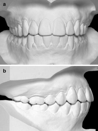
Fig. 8.1
Frontal (a) and profile right (b) views of the dental cast of a subject with optimal dental occlusion. Note that the upper arch is wider than the lower one, there is no space between adjacent teeth (diastema), there is no space between the two arches (normal overbite and absence of open bite), the upper incisors are in front of the lower incisors (normal overjet), the upper and lower dental midlines coincide, and there is no teeth rotation or abnormal teeth inclination
Figure 8.2 shows the three basic sagittal relationships between the incisors (overjet). In the case of ideal overjet (Fig. 8.2a), the upper incisors are in front of the lower ones with little or no space between them. The increased overjet (Fig. 8.2b) consists of an excessive horizontal distance between the teeth, whereas the reverse overjet (Fig. 8.2c) requires that the lower teeth are in front of the upper ones.
Fig. 8.2
Sagittal relationship between the incisors. Ideal overjet with the upper teeth in front of the lower ones (a), increased overjet with excessive horizontal distance between the teeth (b), reverse overjet with the lower teeth in front of the upper ones (c)
The sagittal relationships between the dental arches can be divided into three main classes. Class I presents the ideal posteroanterior relationship between the two dental arches (Fig. 8.3a). Class II is a too anterior upper dental arch and/or a too posterior lower dental arch (Fig. 8.3b). Class III is a too posterior upper dental arch and/or a too anterior lower dental arch (Fig. 8.3c).
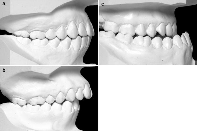
Fig. 8.3
Sagittal classification of dental occlusion. Class I: ideal posteroanterior relationship between the two dental arches (a). Class II: too anterior upper dental arch and/or too posterior lower dental arch (b). Class III: too posterior upper dental arch and/or too anterior lower dental arch (c)
Figure 8.4 shows the three basic vertical relationships between the upper and lower incisors (overbite). In the case of ideal overbite, the upper incisors are in front of the lower ones with a limited overlap between them of 1–2 mm (Fig. 8.4a). The deep bite consists of an excessive overlap between the teeth (Fig. 8.4b), whereas the open bite (Fig. 8.4c) requires the presence of a vertical separation between the teeth.
Fig. 8.4
Vertical relationships between the upper and lower incisors. Ideal overbite with a limited overlap of 1–2 mm between the incisors (a). Deep bite or excessive overlapping between the incisors (b). Open bite or excessive vertical distance between the margins of the incisors (c)
Assessment of the occlusal transversal relationships considers the presence of a midline deviation between the dental arches, a crossbite between opposite teeth, and/or a cant of the occlusal plane (Fig. 8.5).
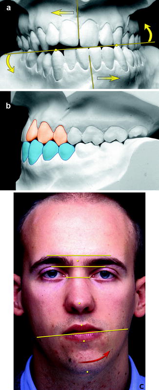
Fig. 8.5
Dental malocclusion with mandibular deviation on the left cant of the occlusal plane (a), dental crossbite of some teeth (b) associated with facial asymmetry and cant of the lip commissures (c)
One important task of the examiner is to correlate the intraoral findings with the external facial appearance of the subject in order to establish the role of dental occlusion on the facial aesthetics. The long-term support effect of particular teeth on the lips and cheeks is also considered. With aging, the soft tissue envelope aesthetic is progressively more dependent on the skeletal support offered by well-aligned and correctly inclined frontal teeth and the surrounding alveolar bone (Fig. 8.6).
Fig. 8.6
The tracings show the intimate relationship between the inclination of the upper incisors and the upper lip position
8.2 Upper Frontal Teeth Assessment
The upper frontal teeth need special attention in all patients, even if they are not mentioned in the patient priority list. Their role in overall facial aesthetics is so important that a qualitative and sometimes quantitative analysis can add many details that help in planning treatment. The upper frontal teeth consist of the four incisors, the two canines, and the four premolars; these teeth should occupy the larger part of the smile. The teeth shape, color, alignment, and symmetry must be evaluated, as well as the architecture and color of their gingival margin. Figure 8.7 shows the detail of well-proportioned and aligned upper frontal teeth.
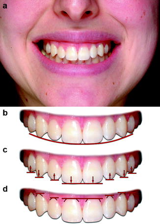
Fig. 8.7
Smiling view of a well-proportioned and aligned upper frontal teeth group (a). It can be noted that the outline created by the occlusal margin forms a gentle curve (b), adjacent teeth have different marginal vertical positions (c), and the gingival margins also have a different shape and position (d)
One of the most difficult and at the same time most important aspects to assess is the anteroposterior projection of the upper teeth relative to the facial profile. The orthodontic literature has called this topic “anterior limit of dentition.” Clinically and using photographs, the full facial profile view at rest must be compared with the same during a posed smile. Figure 8.8 shows the assessment of the anterior projection of the upper incisors in three different clinical cases.
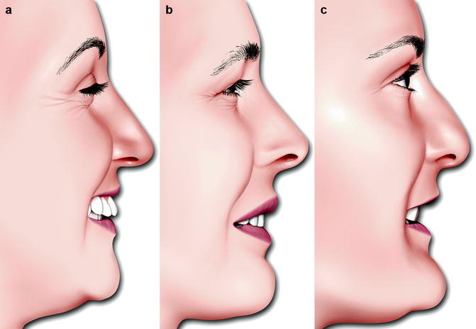
Fig. 8.8
Full facial profile view during smiling. Clinical examples of excessive (a), ideal (b), and reduced (c) sagittal projection of the upper anterior teeth relative to the facial profile
Two particular aesthetic problems of the upper incisors are the midline diastema and the black triangle. The midline diastema is represented by a space between the two upper incisors (Fig. 8.9b), whereas the so-called black triangle is a base-up triangular space bounded by an excessive mesial inclination of the two upper first incisors (Fig. 8.9c). In both cases, the observer and the patient usually perceive the dark shadow between the teeth as an aesthetic problem.
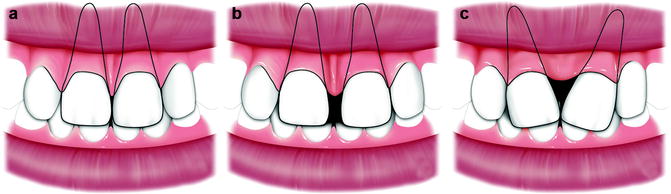
Fig. 8.9
The most favorable aesthetic relationship between the upper incisors, as well as other teeth, requires considerable contact between the two parallel structures (a). The presence of a midline dark shadow between upper central incisors may be a diastema (b) or a “black triangle” (c)
The spatial position and inclination of the incisors are discussed further in Sect. 9.6.
8.3 Lips Assessment
The first problem in the assessment of the lips is to obtain a truly relaxed position from the patient. When reviewing a complete set of photographs of a clinical case, it is not unusual to discover that the lip position is not constant but changes from a complete seal, most frequently at the start of the photographic session, to a more relaxed (habitual!) one with the presence of a more or less wide space between the two lips (Fig. 8.10). A partial nasal obstruction should be suspected when the subject has the habit of maintaining a space between the lips and breathing through the mouth.
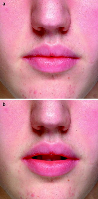
Fig. 8.10
These photographs were taken in sequence during the same sitting without requesting the patient to take a particular lip position. After a few seconds in which the patient maintained a lip seal sustained with some muscular contraction (a), he returned to his habitual relaxed position with loss of contact between the lips (b)
Figure 8.11 shows a case of lip incompetence with absence of lip seal at rest due to an increased overjet with mandibular hypoplasia. In these cases, maintaining perfect lip closure requires a voluntary contraction that results in the flattening of the lip profile curves.
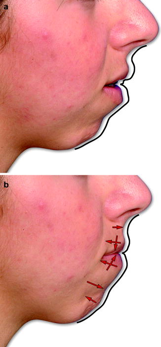
Fig. 8.11
A case of lip incompetence with absence of lip seal at rest due to an increased overjet with mandibular hypoplasia (a). In these cases, maintaining perfect lip closure requires a voluntary contraction that results in the flattening of the lip profile curves (b)
8.3.1 The Attractive Female Lips
Knowing a prototype of younger, “healthy,” and attractive female lips helps us to understand the roles and effects of intrinsic lip problems and any possible correlation with a dentofacial deformity, the aging process, or an acute lip trauma; it also helps to define the younger, “healthy,” and attractive male lips.
Figures 8.12 and 8.13 show this prototype in frontal and profile views, as well as the preferred anatomical terms of the perioral region. The characteristics of attractive female lips can be summarized as follows:
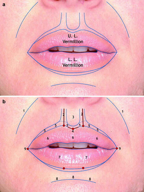
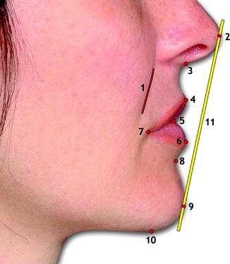
Fig. 8.12
Frontal views of younger, “healthy,” attractive female lips (a, b). Perioral soft tissue determinants (b): 1 nasolabial sulcus, 2 philtrum columns, 3 philtrum, 4 Cupid’s bow, 5 lip white roll, 6 upper lip vermillion (its central and more projecting portion is also called the upper lip tubercle), 7 lower lip vermillion, 8 labiomental sulcus, 9 lip commissures (the distance between the two commissures is defined as mouth width)
Fig. 8.13
Profile view of younger, “healthy,” attractive female lips. Reference points and Ricketts’ lips projection reference E-line: 1 nasolabial sulcus, 2 nasal tip, 3 subnasale, 4 labrale superior, 5 stomion, 6 labrale inferior, 7 lip commissure, 8 labiomental sulcus, 9 soft tissue pogonion, 10 soft tissue menton, 11 Ricketts’ E-line. The series of gentle curves from nasal tip to soft tissue menton characterize the lips profile
Stay updated, free dental videos. Join our Telegram channel
VIDEdental - Online dental courses


