Lip cancer is one of the more common cancers of the head and neck region and should be one of the most curable because of its ability to be detected in the early stages. In general terms, its prognosis is quite favorable, with 5-year survival statistics exceeding 90% in most reports. Some lip cancers have been observed to exhibit aggressive behavior, however, with recurrence or mortality noted in up to 15% of cases. Lymph node metastases seem to occur in 5% to 20% of patients, and the overall incidence is quoted at 10%. The frequency of lip cancer varies internationally, with an incidence of 30% of all cases of oral cancer in certain regions. In the United States the incidence of lip cancer is 1.8 per 100,000 population.
Lip cancer is occasionally described as being one of the oral cancers. These authors choose to consider lip cancer as one of the facial skin cancers for anatomic and logistic reasons. Anatomically, the most common location for lip cancer is the vermilion or mucocutaneous junction. In general, the behavior of cancer of the lip is similar to that of skin cancer rather than oral mucosal cancer in terms of both survival and lymph node metastases. Finally, the most frequently identified carcinogen of the most common lower lip cancer, squamous cell carcinoma, is ultraviolet radiation, as is the case for the most common type of upper lip cancer, basal cell carcinoma. For these reasons, lip cancer should be considered a skin cancer rather than an oral cancer.
Approximately 90% of cases of lip cancer occur on the lower lip, with nearly 7% of cases occurring on the upper lip and the remainder located at the commissure. Squamous cell carcinoma is clearly the most common histologic variant, with basal cell carcinoma, melanoma, and minor salivary gland cancers of mucosal origin constituting the other histologic variants. The latter diagnosis is certainly an exception to the previous statement that lip cancer should be considered a skin cancer rather than an oral cancer. When they occur, minor salivary gland neoplasms are most commonly seen within the mucosa of the upper lip, and the overwhelming majority of these tumors are benign. Approximately 95% of lip cancers occur in men, typically those older than 50 years. The most common age range at diagnosis is 54 to 65 years, although these cancers will occasionally occur in patients younger than 30 years.
Etiopathogenesis/Causative Factors
The etiology of lip cancer is probably best described as multifactorial and perhaps poorly understood, as is the case with many human cancers. Approximately a third of lip cancers are associated with excessive sun exposure in patients with outdoor occupations. Most tumors originate on the exposed vermilion of the lower lip. Lip cancer is seen commonly in patients with second primary skin malignancies. Like other head and neck skin cancers, these patients may have light complexions, freckles, blue eyes, and fair-colored hair. The lower lip is at higher risk for skin cancer than the upper lip because of the prominence of the lower lip. This anatomic feature accounts for the discrepancy between the incidence of upper and lower lip cancers. The prevalence of lip cancer is at least 10 times higher in whites than in those with darker skin, and it is very rare in black people.
Other risk factors for lip cancer include the traditional carcinogens for head and neck cancer such as cigarette and pipe smoking, lip trauma, and immunosuppression. Although these risk factors may be related to the development of lip cancer, the overwhelming anecdotal evidence points to prolonged and cumulative sun exposure as the most significant carcinogen involved in the development of melanoma and non-melanoma lip cancers.
Pathologic Anatomy
The upper lip is formed embryologically by fusion of the lateral maxillary processes and a central nasofrontal process. Because of anatomic separation of the lateral segments, contralateral lymphatic cervical metastases are quite rare from upper lip cancers. This anatomic feature is in contradistinction to the lower lip, which forms by the fusion of two lateral mandibular processes in the midline. Lower lip cancers, therefore, are at higher risk for contralateral metastasis. The blood supply to the lips is derived from the superior and inferior labial arteries.
Lymphatic drainage of the lips follows a predictable course of metastatic dissemination. Lymph node metastases related to lip cancer occur in fewer than 10% of patients with cancer of the lower lip and in up to 20% of patients with cancer of the upper lip and commissure. Cancers of the lateral aspect of the upper lip preferentially metastasize to the buccal, periparotid, and preauricular region overlying the body of the mandible. Secondary metastases will occur to the cervical lymph nodes in the submandibular triangle. Cancers of the lower lip preferentially drain into the cervical lymph nodes of the submental and submandibular triangles of level I cervical lymph nodes. Subsequent metastases can occur in level II and III lymph nodes. Dissemination to level IV and V lymph nodes is quite rare, although when cervical metastases do occur from lower lip cancers, bilateral level I metastases are not uncommon.
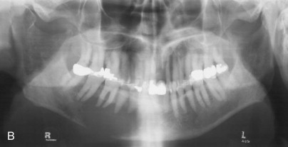
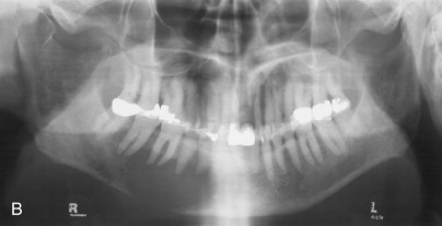
The sensory nerve distribution of the upper and lower lips is provided by the maxillary and mandibular divisions of the trigeminal nerve, respectively. The neurosensory innervation of the lower lip is of significance in the evaluation of patients with lower lip cancer, specifically with regard to obtaining a screening panoramic radiograph to rule out mandibular involvement by the cancer as a result of perineural spread ( Fig. 63-1 ).
Diagnostic Studies
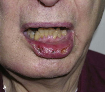
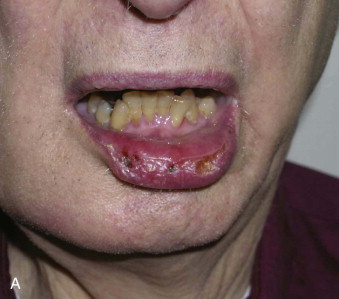
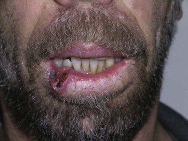
The history, physical examination, and incisional biopsy are the most valuable tools in establishing a diagnosis of lip cancer. A history of a non-healing crusted lesion of the lip that persists for several months is typical. Physical examination will reveal an area of crusting and surrounding induration or a mass, depending on the chronicity of the cancer ( Fig. 63-2 ). The incisional biopsy should be performed within the center of the lesion to establish the diagnosis. Once the diagnosis has been made, it is most important to obtain a panoramic radiograph to investigate for a widened mental foramen or erosion of the mandible, which could occur by perineural spread of a lower lip cancer (see Fig. 63-1 ). In general terms, special imaging studies such as computed tomography (CT), magnetic resonance imaging, and positron emission tomography (PET/CT) are not required to assist in ablative surgery associated with a lip cancer when clinical neck examination does not reveal suspicious adenopathy (N0). When the neck is classified as N+, however, special imaging studies, particularly PET/CT, are indicated. This is especially the case when unilateral adenopathy exists in association with a lower lip cancer and PET/CT will provide images of the contralateral neck. Distant metastases are identified in less than 2% of patients at the time of initial evaluation of a previously untreated lip carcinoma. In preparation for ablative surgery performed in an operating room setting, routine blood work, an electrocardiogram, and chest radiographs are obtained according to standards set by the surgeon’s operating room and attending anesthesiologists.
Treatment/Reconstructive Goals
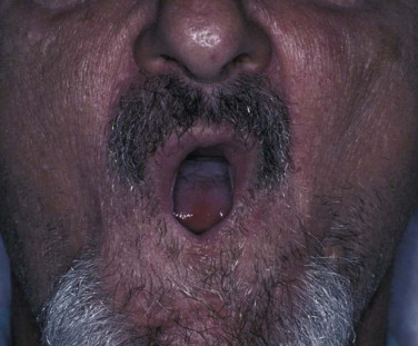
Management of lip cancer should include the objectives of proper ablation of the patient’s cancer and functionally and esthetically acceptable immediate biologic reconstruction of the lip. To this end, lip cancer is unique in that there is no ability to negotiate for delayed biologic reconstruction. This statement represents a departure from the management of some head and neck cancers, where alloplastic reconstruction of the mandible with a bone plate can take place while performing delayed biologic reconstructive surgery of a segmental defect. Lip cancer must be managed with immediate soft tissue reconstruction. In so doing, the reconstruction must be performed in a manner that avoids microstomia ( Fig. 63-3 ). Soft tissue flaps, as may be used in these reconstructions, may be categorized according to their blood supply. Three flap patterns are recognized: random-pattern, axial-pattern, and microvascular free flaps. Random-pattern flaps are those in which specific pedicles are not identified or necessarily preserved within the flap. By contrast, an axial-pattern flap is one in which the pedicle is identified and intentionally preserved within the flap that is rotated into the recipient tissue bed. Axial-pattern flaps for head and neck reconstruction may be local or regional according to their anatomic site of origin, whereas axial- and random-pattern flaps for lip reconstruction are distinctly local in nature. Finally, microvascular flaps involve distant soft tissue transfers in which arterial and venous anastomoses are required for flap viability.
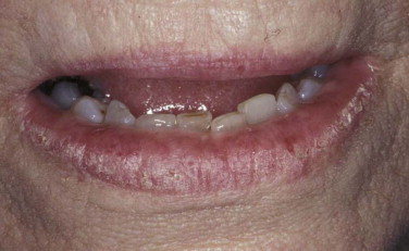
Any discussion of cancer undoubtedly warrants a discussion of pre-cancer. The most common pre-cancer of the lower lip is actinic keratosis ( Fig. 63-4 ). Actinic keratosis results from damage to the lower lip by ultraviolet radiation. It is named for the white color of the vermilion of the lower lip that is affected by actinic keratosis. The lip is also characteristically dry in its appearance. Ulcerations may be present, but a mass is not noted in patients with actinic keratosis. Histologic evaluation of actinic keratosis may reveal signs of dysplasia or carcinoma in situ. When a mass is present, a diagnosis of invasive cancer is almost certain to be established through the required incisional biopsy. Actinic keratosis is a clinical diagnosis that does not require preoperative incisional biopsy but certainly necessitates histologic confirmation at the time of excision.
Stay updated, free dental videos. Join our Telegram channel

VIDEdental - Online dental courses


