Fine-Needle Aspiration Cytology and Histological Biopsies
Magnetic Resonance Imaging and Computed Tomography
Preparation and Scheduling of Surgery
Introduction
Statistically, most salivary gland tumors are benign. A standardized approach to a patient with a salivary gland lump will help minimize the risk of overlooking malignant disease. This standardized approach also helps reduce the risk of suddenly finding a benign tumor in a more unconventional location during surgery, or tumor extension into an unfamiliar anatomic region, or unexpectedly requiring a different surgical approach for which the patient’s consent should have been obtained beforehand. This chapter outlines a standard, step-by-step sequence of investigations for patients with benign salivary gland tumors (Fig. 21.1).
Clinical Features
Patients typically report the incidental appearance of a lump in the parotid or submandibular region. The tumor may have been present for a long period, with a slow growth rate. An intraoral tumor in the minor salivary glands or a tumor in the deep lobe of the parotid gland may first be detected accidentally by a dentist or physician, or occasionally when the tumor reaches a more advanced stage or size at which it starts to interfere with swallowing or speech functions. Benign salivary gland tumors mainly present as painless swellings of the affected gland (for more details on the differential diagnosis, see Chapter 6).
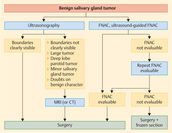
Fig. 21.1 Investigation algorithm for benign salivary gland tumors.
 Most malignant tumors may also present as a painless swelling of a salivary gland; the symptom does not rule out malignancy.
Diagnostic Work-Up
 Clinical Examination
On examination, a benign tumor of the parotid or submandibular gland usually appears as a well-defined, nontender, mobile mass. Additional clinical signs such as skin involvement or attachment, associated nerve palsy, or fixation to underlying structures are signs suggestive of malignancy. In the parotid, most benign tumors are located in the lower third of the gland (the “tail”), but the tumor can appear anywhere in the gland. Larger size, a lack of clear demarcation, or a lack of mobility may suggest that the tumor originates in or extends into the deep lobe of the parotid gland. Benign tumors of the minor gland are rare. The most common sites involved include the palate, parapharyngeal space, and lacrimal gland. Due to the lack of surrounding soft tissue and absence of a capsule, benign tumors of the minor salivary gland often appear to be more fixed than mobile. It should be borne in mind that the majority of tumors of the minor salivary glands are not benign but malignant. A salivary gland tumor in the parapharyngeal space may present initially only with medial displacement of the soft palate and/or tonsil. In these cases, palpation of the tonsil is very helpful for defining the margins of the tumor.
 Examination not only of the primarily affected salivary gland, but also of all major glands and other areas in which minor salivary glands are located is necessary in order to exclude the synchronous appearance of salivary gland tumors at the other sites in the head and neck region.
< div class='tao-gold-member'>
Stay updated, free dental videos. Join our Telegram channel
VIDEdental - Online dental courses


