Fig. 3.1
Roadmap of comprehensive patient evaluation in esthetic dentistry
For the initial visit of the patient, a private consultation room is advised, as this allows the meeting to take place in a quiet, personalized and relaxing environment in which the dentist can learn the desires, concerns, and expectations of the patient alongside the dental and medical history and determine the current situation [7]. This personal interview also lends credence to the patient’s first impression of the clinician and clinical environment, and hence can create a positive clinician–patient relationship [6]. The collected data at this initial conversation are also mandatory for developing different treatment approaches for the subsequent course of treatment and ultimate outcome. During the second appointment, arranged to last much longer, the clinician should obtain specific information, perform examinations, and discuss the several possible treatment approaches with the patient. Sufficient time should be allotted at this stage to gather information and specific data on dental history, to perform diagnostic tests and to consult with medical specialists if necessary.
3.1 Medical History
For documenting the medical history of patients a standard, detailed history form should be completed at this stage to gather specific information to help identify any conditions that could contraindicate, complicate, or alter the treatment procedure [8]. With this detailed form, clinicians can obtain information on infections or systemic disease that may require specific prevention, prophylactic therapies, treatment modifications, and referral to medical specialists. Allergic reactions to drugs and restorative materials can contraindicate the restorative procedures, and physiological changes due to aging may affect the treatment plan. Besides such information, clinicians can also learn about the habits of the patients such as smoking, plaque control, and other pernicious habits (i.e., thumb sucking). Dietary habits of the patients also need to be recorded at this stage for both present and future restorative and oral care. Knowledge of previous dental visits and treatments is also essential for gathering information about past dental problems, previous conditions, and patient responses to procedures, to provide optimum oral care for future treatment plans [8]. In addition, a history of previous orthodontic therapy is important when planning the treatment because this may indicate the tendency of diastema to recur. All of this information must be documented scrupulously on the history form, to be used as needed.
3.2 Clinical Examination
After recording all necessary information on the history form, the next step should be detailed clinical evaluation of all determinants of the mouth such as periodontal condition, general tooth health, oral soft tissue, occlusion, and extraoral examination. As these patients are already seeking esthetic restorative treatment, a comprehensive clinical esthetic examination by listening sufficiently to the patient’s desires and expectations is also mandatory for a satisfactory outcome [9].
3.3 Extraoral Examination
Although generally facial symmetry, lip position at rest and at smile, facial contours, and midline axis may not be major evaluation criteria in simple or routine restorative treatment procedures, all of these are important components of esthetic restorative procedures [7]. Determination of the harmonious function or dysfunction and all components of the stomatognathic system should be carefully evaluated step by step. For evaluation of the mandibular movement and switching limits, the clinician should be positioned at the 12 o’clock position (Fig. 3.2) to determine minor deviations during these motions. In addition, this position can allow the clinician to evaluate the pain response of the patient by pressing onto the capsule during the switching motion. Temporomandibular joint (TMJ) examination by palpation can also be performed at the 12 o’clock position (Fig. 3.3). Even in the absence of any significant symptom, it is important to record mandibular motion, switching limits, mastication, and the comfort of the patient during the extraoral evaluation of these movements [7, 10]. Significant TMJ changes (i.e., intracapsular) may occur with no symptoms, which could affect the long-term outcome of restoration and occlusion [10]. For this reason, early evaluation of the stomatognathic system with all its components is important. The information gathered at the initial conversation also allows a more helpful examination at this stage, as patients may report concerns with TMJ problems, rattling noise during chewing, headaches, or muscle tension [10]. Clicking noises can be due to a disk displacement [7, 11], and may require a detailed evaluation and physical therapy by a clinical expert in the field.
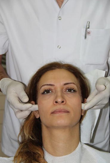
Fig. 3.2
Evaluation of the patient from 12 o’clock position
Fig. 3.3
Temporomandibular joint (TMJ) examination at 12 o’clock position by palpating onto the disks
Key Note
Examination of all components of the stomatognathic system at the beginning is important for long-term stability and success.
Palpation of the TMJ can be done when the mandible is closed and opened widely (Fig. 3.4a, b). At the closed position of the mandible, one can evaluate the lateral pole and interrelated surrounding structures by palpating. When the mandible is widely opened, condylar movement occurs downward and forward throughout the articular eminence, and at this position the posterior part of the TMJ can be palpated [10]. During these palpations, patients with healthy structures generally should not feel any pain or discomfort. Any discomfort or pain detected during the TMJ palpation could be due to dropsy or inflammation in those structures, probably caused by trauma such as bruxism, malocclusion, or any kind of facial accident history [10]. Evaluation of mandibular motion is another important criterion whereby maximum opening, lateral and protrusive movement of the mandible should be measured for consideration. In all these mandibular movements there should be no pain or discomfort, and if so it should be specifically detected through evaluating the motion while the patient is talking to specify the area of discomfort. These pains or discomforts may be TMJ induced, muscle related, or both. Normally, approximately a 50 mm opening and 10 mm excursive movements should be seen on mandibular motion [10]. Any deflection in these average limits detected during examination might suggest problems with the TMJ, especially with the capsular structures and muscles. Another important test that should be borne in mind for testing the TMJ is the superior compression test, whereby bimanual guidance is used to measure the load-bearing capacity of the TMJ [10] (Fig. 3.5). If pain and tension are apparent during this test, they probably arise in various forms from intracapsular or surrounding structures [10]. If the patient feels pain or tension during the superior compression test and points out a location in the TMJ area, this generally indicates intracapsular problems [10]. If tension is reported by the patient during the bimanual superior compression test, this may suggest hypercontraction of the lateral pterygoid muscles [10], which pulls pterygoids down and forward through the articular eminence. If sensitivity is reported by the patient during the bimanual compression test, this would indicate inflammation or dropsy in the capsular area [10]. Besides palpation, clinicians can also use auscultation devices such as ultrasonography to detect the current condition and any TMJ problems [10]. A step-by-step evaluation chart for all the components of the TMJ can be helpful for acquiring the necessary information to be kept for future consultation when needed. When these signs or conditions are detected by either palpation or auscultation during the extraoral examination of the patient, it must be definitively treated before any restorative treatment procedure to achieve a long-term satisfactory result.
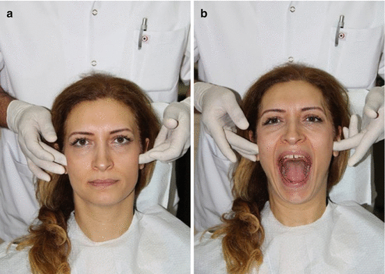
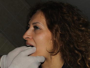
Fig. 3.4
Palpation of the TMJ (a) when mandible is closed and (b) mandible is opened widely
Fig. 3.5
Load-bearing capacity examination of the TMJ using bimanual guidance
3.4 Examination of Muscles
Testing the masticatory muscles is another important extraoral examination, for which various kinds of muscle examination such as palpation, the patient’s own statement during function, and provocation tests are available [10]. While palpating, normally healthy and well-functioning muscle should not exhibit any hypertrophy, sign, or pain during examination. Any discomfort or pain detected on palpation would suggest that muscle is in hypercondition or hypertrophy. A variety of reasons can provoke muscle discomfort or pain, such as a pathological condition (e.g., atypical facial pain, migraine), parafunctional conditions (clenching, grinding, bruxism), and musculoskeletal disorders [7, 10]. Occlusal instability and bruxism can provoke muscle discomfort during function especially on the excursive movement of the posterior teeth, and hence creates pain by hyperactivating the elevator muscles [10, 12, 13]. In general, extraoral muscles are evaluated first, followed by the intraoral muscles. During extraoral examination the masseter muscle, temporalis muscles, occipital muscle, sternocleidomastoid muscles, medial pterygoid muscle, and digastric muscles should be palpated in sequence [7, 10]. When palpating these muscles, the patient is asked to point out exactly where a pain or discomfort occurs. The masseter and temporalis muscles are examined by palpation primarily at rest (Fig. 3.6a, b); the patient is then asked to clench the jaw (Fig. 3.7a, b) to allow evaluation of any differences detected between these palpations such as hardness on these muscles during this function [7, 14]. If any differences are detected during the evaluation, the patient is asked about any parafunctional habits such as nighttime bruxism, clenching, or any history of chronic headache [7]. Painful response or discomfort of patients during palpation of these muscles may also contribute to non-dental problems such as migraines and atypical facial pain; hence, a true-positive evaluation is mandatory [7]. During examination of occipital muscles (Fig. 3.8), if the patient gives a positive response to palpation this may signify a possible cervical component problem related to the discomfort, and before initiation of any restorative procedure the patient must be referred to a specialist for a full evaluation of TMJ and cervical muscle [7, 15]. When evaluating the sternocleidomastoid muscle by palpation (Fig. 3.9), the patient may refer to pain at the masseter and TMJ areas; in this case a detailed careful examination of all areas should be carried out [7]. In addition to the extraoral examination, an intraoral examination of medial pterygoid muscles (Fig. 3.10), the reference area of which is pterygomandibular groove, is important. If the patient reports any discomfort or sensitivity during palpation of this muscle, it may generally be related to posterior excursive interference [10]. The lateral pterygoid (Fig. 3.11) is another important muscle that is difficult to examine directly intraorally, owing to its anatomical location [7, 10]. Positioning in front of the patient, the clinician places his or her thumbs on the tip of the chin and asks the patient to move the mandible forward. During this motion more resistance will be generated by the thumb and the patient may feel discomfort or pain if there is a problem with the muscle at the anterior region of the condyle [10]. However, it must be borne in mind that due to the extended time period afforded by this examination, lactic acid can be deposited into the muscle even in healthy mouths, and may become painful for the patient [10]. Lastly, during palpation of the digastric muscle (Fig. 3.12) which is predominantly responsible for the opening of the mouth, patients will usually report pain or discomfort if there is any dysfunction of the muscle [7, 16]. In general, healthy muscles do not generate any pain and discomfort during palpation [17]. The importance of muscle examination to both the restorative treatment procedure and the restorations must be clearly explained to the patient. If there are any signs of discomfort or pain during the muscle examination or via self-reporting by the patient, esthetic restorative treatment should be postponed until full TMJ and muscle therapy is completed by an expert in the field [7].
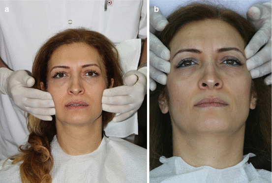
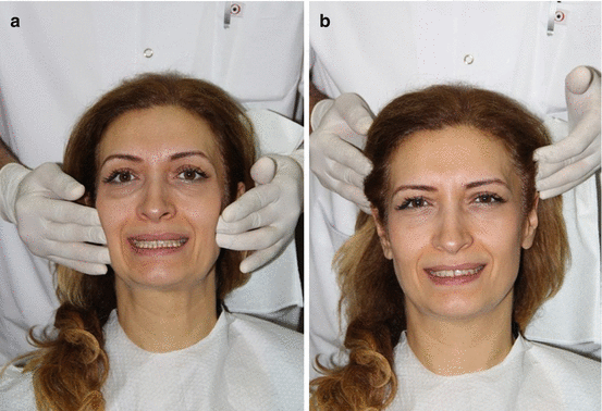
Fig. 3.6
(a) Palpation of masseter muscle at rest. (b) Palpation of the anterior temporalis at rest
Fig. 3.7
(a) Palpation of masseter muscle while clenching. (b) Palpation of the anterior temporalis muscle while clenching
Stay updated, free dental videos. Join our Telegram channel
VIDEdental - Online dental courses


