Introduction
Orthodontists are often asked to remove fixed retainers before magnetic resonance imaging (MRI). This study was undertaken to assess the effects of 2 commonly used fixed retainers on MRI distortion and whether they should be removed.
Methods
MRI scans were performed on a dry skull with Twistflex (Dentaurum, Ispringen, Germany) and Ortho Flex Tech (Reliance Orthodontic Products, Itasca, Ill) retainers. Two neuroradiologists independently ranked the distortions. The influence of the fixed retainers’ alloys, their distance to the area of diagnosis, location, strength of the magnetic field, and the spin-echo sequence were examined. Statistical analysis included kappa and Pearson chi-square tests.
Results
Ortho Flex Tech retainers caused no distortion. Twistflex retainers caused distortion in 46% of the tests in areas close to the retainer (tongue and jaws). Maxillary fixed retainers and the combination of maxillary and mandibular fixed retainers further increased the distortion. Greater distortion was observed with 3-T magnetic fields and T1-weighted spin-echo sequences.
Conclusions
Removal of the Ortho Flex Tech retainer is unnecessary before MRI. Removal of the Twistflex should be considered if the MRI scans are performed to diagnose areas close to the fixed retainers, when 3-T magnetic fields and T1-weighted sequences are used, and when both maxillary and mandibular fixed retainers are present.
Highlights
- •
Removal of fixed orthodontic retainers is not necessary before MRI scans for regions distant from the retainer.
- •
Ortho Flex Tech retainer should not be removed prior to MRI scanning.
- •
Twistflex retainers should be removed for MRI scans of regions close to the retainer.
- •
The presence of both maxillary and mandibular retainers increases the distortion.
Magnetic resonance imaging (MRI) has become a common diagnostic tool for various medical and dental conditions (migraine and cluster headaches, epilepsy and other seizure disorders, multiple sclerosis, head and neck tumors, temporomandibular joint disorder, and many others). The advantages of MRI over other imaging techniques include excellent soft-tissue quality and absence of ionizing radiation. The common MRI apparatus, often designated by their magnetic fields, are of 1.5 and 3 teslas (T), which represent the magnetic intensity of the machine. Ferromagnetic materials (several common metals) distort the magnetic field and result in distortion of the magnetic resonance image, affecting its diagnostic value.
Recently, there has been an increase in the number of orthodontic patients worldwide. According to Proffit et al in 1998, over 30% of white youths, 11% of Mexican-Americans, and 8% of African Americans reported receiving orthodontic treatment. At the end of active orthodontic treatment, the results are usually retained for many years using fixed retainers made of a metal wire bonded to the lingual side of the anterior teeth.
Owing to the increased number of MRI referrals, orthodontists are frequently requested to remove metal orthodontic appliances before the MRI procedures. Removal and reinstallation of an orthodontic appliance is laborious and bears a risk of relapse of teeth to undesired positions. As a result, orthodontists are interested in understanding the effects of orthodontic appliances on MRI imaging to minimize the need for removal.
The presence of orthodontic appliances has 3 important implications for MRI diagnosis. First, magnetic field interactions may pose a risk of detachment to the patient. Loose orthodontic devices pose a significant danger for the patient. Second, metals in the appliances may cause heating. Third, they may produce image distortions that could affect the diagnostic quality of the MRI.
Only a few studies have been conducted to evaluate the influence of different fixed orthodontic appliances on the diagnostic quality of magnetic resonance images. One study showed that stainless steel brackets cause significant distortion, rendering several cranial regions nondiagnostic, whereas plastic, ceramic, and titanium brackets cause minimal interference.
The effect of different types of fixed retainers has scarcely been addressed so far. When bonded firmly, fixed retainers pose no risk to the patient in the magnetic field of a clinical MRI scanner. However, image quality may be significantly affected.
The purpose of this study was to assess the in-vitro effects of the 2 most common types of fixed retainers on MRI distortion and answer the dilemma of whether they should be removed before MRI.
Material and methods
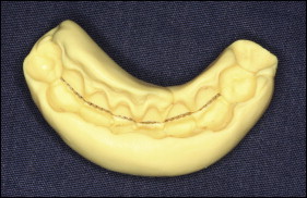
A dry skull of a female was used for this study. Alginate impressions of the maxillary and mandibular dental arches were taken, and plaster models were poured. The 2 types of fixed retainers were fabricated directly on the lingual aspects of the maxillary and mandibular incisors and canines of the plaster models. The fixed retainer wire was attached to the lingual surface of each tooth using dental floss. Polyvinylsiloxane key (Elite HD + type 0 putty consistency; Zhermack, Badia Polesine, Italy) was prepared for each jaw to hold the retainer in place. The retainers were embedded into this material ( Fig 1 ).
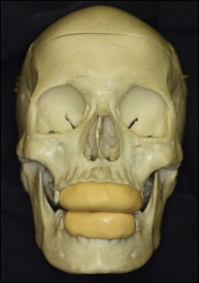
For each retainer key, the skull was immersed in a container including a diluted 2% solution of gadolinium contrast agent (Dotarem; Guerbet, Villepinte, France) and placed in the scanner. The MRI sequences were performed for that particular set; then a different tray was inserted, and subsequent magnetic resonance scans were completed ( Fig 2 ). The diluted gadolinium solution created a background signal to the bone that simulated that of soft tissues. It did not affect the metal distortion artifacts but allowed them to be seen using MRI sequences identical to those in clinical imaging.
To determine the effect of the polyvinylsiloxane key on the MRI scans, 2 scans were carried out on the key without the retainers, which served as the controls.
Two experienced neuroradiologists (J.M.G. and E.B.-D.) independently evaluated the images to determine the amount of distortion. All images were presented at once on the neuroradiologists’ computer screens. The images were randomly labeled. The neuroradiologists were asked to rank the images according to the distortions in these regions using a modified receiver operating characteristic method of distortion classification ( Table I ).
| Score | Image appearance | Diagnostic or nondiagnostic |
|---|---|---|
| 1 | No distortion or artifact | Diagnostic |
| 2 | Minimal distortion or artifact | Diagnostic |
| 3 | Moderate distortion or artifact | Moderately diagnostic |
| 4 | Severe distortion | Nondiagnostic |
The following 5 variables were examined.
- 1.
The type of alloy of the fixed retainer. Two commonly used fixed retainers were chosen for this study: Twistflex, 0.18 in (Dentaflex, triple strand twisted; Dentaurum, Ispringen, Germany), an alloy of stainless steel (chromium, 17%-20%; nickel, 8%-12%; carbon, 0.08%-0.15%; the rest is mainly iron); and Ortho Flex Tech (Reliance Orthodontic Products, Itasca, Ill), an alloy composed of gold, 58.33%-58.70%; silver, 1.5%-6.0%; copper, 28.0%-31.0%; nickel, 0.3%-6.5%; and zinc, 5.0%-6.3%.
- 2.
The distance between the region of interest and the retainer. Ten regions of the head were assessed: tongue, mandible, anterior maxilla, posterior maxilla, orbits, nasopharynx, pituitary gland, frontal lobe, temporal lobes, and brain stem.
- 3.
The location of the fixed retainer. Six trays were fabricated: maxillary and mandibular Twistflex, maxillary and mandibular Ortho Flex Tech, and maxillary and mandibular control polyvinylsiloxane keys without retainers.
- 4.
The strength of the magnetic field. The scans were performed in the MRI unit, Hadassah Medical Center, Jerusalem, Israel, on both 1.5-T (Avanto) and 3-T (Trio) systems (both, Siemens Medical Solutions, Erlangen, Germany) using standard multi-element head coils.
- 5.
The spin-echo sequence. Imaging sequences included axial fast-spin echo T2-weighted images (TR/TE 3500/90 ms), axial and sagittal conventional spin-echo T1-weighted images (TR/TE 500/14 ms), with a slice thickness of 5 mm and a 20% gap.
Statistical analysis
Statistical analysis included descriptive statistics, kappa tests to evaluate interexaminer calibrations, and Pearson chi-square tests. The Pearson chi-square test was used to compare the distortions caused by the different retainers and different MRI settings. The significance level for the statistical testing was set at P <0.05. The analysis was carried out using SPSS software (version 21.0; IBM, Armonk, NY).
Results
Statistically significant differences were found between the distortion scores assigned to the Twistflex and Ortho Flex Tech retainers ( P <0.001). No distortion was observed with the Ortho Flex Tech retainers, similar to the control, whereas the Twistflex retainers caused distortion in 46% of the tests. In 20% of the cases, the distortion was minimal; in 7%, it was moderate; and in 19%, it was severe ( Table II ).
| Wires | Distortion | Total | |||
|---|---|---|---|---|---|
| None | Minimal | Moderate | Severe | ||
| TF | |||||
| Count | 54 | 20 | 7 | 19 | 100 |
| % | 54.0 | 20.0 | 7.0 | 19.0 | 100.0 |
| OF | |||||
| Count | 80 | 0 | 0 | 0 | 80 |
| % | 100.0 | 0.0 | 0.0 | 0.0 | 100.0 |
| Control | |||||
| Count | 20 | 0 | 0 | 0 | 20 |
| % | 100.0 | 0.0 | 0.0 | 0.0 | 100.0 |
The Twistflex retainers mainly affected anatomic regions in their vicinity, such as tongue, mandible, and maxilla. Other anatomic regions, not near the Twistflex retainer, were unaffected ( Table III ). The differences between the distortion scores in the different anatomic regions were highly statistically significant ( P <0.001).
| Distortion | ||||
|---|---|---|---|---|
| None | Minimal | Moderate | Severe | |
| Tongue | ||||
| Count | 26 | 6 | 3 | 5 |
| % in anatomic site | 65.0 | 15.0 | 7.5 | 12.5 |
| Mandible | ||||
| Count | 27 | 11 | 0 | 2 |
| % in anatomic site | 67.5 | 27.5 | 0.0 | 5.0 |
| Maxilla, anterior | ||||
| Count | 23 | 1 | 4 | 12 |
| % in anatomic site | 57.5 | 2.5 | 10.0 | 30.0 |
| Maxilla, posterior | ||||
| Count | 38 | 2 | 0 | 0 |
| % in anatomic site | 95.0 | 5.0 | 0.0 | 0 |
| Orbit and other areas | ||||
| Count | 40 | 0 | 0 | 0 |
| % in anatomic site | 100.0 | 0.0 | 0.0 | 0 |
Stay updated, free dental videos. Join our Telegram channel
VIDEdental - Online dental courses


