Introduction
The Invisalign (Align Technology, Santa Clara, Calif) force delivery system has not been investigated. Since forces are related to the strains developed on the aligner surface, the behavior of the maxillary incisor and premolar von Mises strains (IVM, PVM) were studied during biweekly wear of an aligner.
Methods
Maxillary aligners (n = 61) were examined from 3 patients requiring maxillary incisor retraction and stationary anchored premolars. Two series of maxillary aligners were manufactured. Series 1 was worn by the patient, and series 2 served for in-vivo measurements with 2 strain gauge rosettes bonded to each aligner on the vestibular sides of the incisor and the premolar. Measurements were taken at days 1, 2, 9, and 15.
Results
All aligners demonstrated a peak IVM strain at day 1 ( P <0.001); it then decreased at day 2 and plateaued through day 15. No anchorage loss was found in 2 patients (IVM ≥PVM), and a minute loss was evident in 1 patient (PVM >IVM).
Conclusions
Each aligner should be worn close to 24 hours in the first 2 days, with the time subsequently reduced (remaining 12 days). Final aligners should be thicker or worn for longer period (eg, 3 weeks). In spite of the inherent anchorage property of the aligner, attachment reinforcement should be considered in demanding anchorage requirements.
Orthodontic treatment with Invisalign aligners (Align Technology, Santa Clara, Calif) was introduced in 1999. This system is based on clear sequential appliances (aligners) made of a translucent thermoplastic material. The aligners are worn on a biweekly regimen, and a progressive alignment of 0.2-mm translation or 1° rotation per tooth is designed in each aligner. Three stages precede aligner manufacturing. These include scanning the dental impression, preparing a treatment plan, and manufacturing the aligners. The latter is divided into 2 substages. In the first stage, the patient’s polyvinyl siloxane dental impression is scanned directly with a FlashCT (HYTEC Inc, Los Alamos, New Mexico) to create a 3-dimensional full-volume model. FlashCT is an advanced industrial computed tomography system for high-speed 3-dimensional imaging. In the second stage, geometric modifications of the setups are planned by using a computer-aided design system. An incremental 3-dimensional correction of the malaligned teeth is designed. In the first substage of the third stage, stereolithography (SLA) molds (setups of both dental arches) are fabricated by using SLA technology and computer-aided manufacturing systems. Stereolithography technology is a solid imaging process that uses a laser beam to expose and solidify successive layers of a photosensitive liquid until the desired mold is formed in acrylic resin. In the second substage, the custom-made aligners are formed from a polyurethane resin sheets (polyurethane from methylene diphenyl diisocyanate and 1,6-hexanedial, additives) by using a vacuum thermoforming process in which the resin sheet is stretched over each of the SLA molds.
The removable aligners offer esthetic advantages, as well as oral hygiene maintenance, thus reducing the risks of decalcification, caries, gingivitis, and periodontal disease. The clinical efficiency includes evaluating interproximal reduction, attachments, space closure after extraction, and space distribution for prosthetic restorations. Recently, even complex interdisciplinary managements have been examined—eg, orthognathic approach and soft-tissue correction.
Few studies have considered the biomechanical questions regarding this technology. Greater stiffness and surface modifications were found in the aligners after usage. Ryokawa et al studied the mechanical property of the aligner material and found no significant change in elastic modulus when the Invisalign polyurethane resin sheet was compared before and after thermoforming, and after immersion in water at 37°C. However, Eliades and Bourauel reported that, although Invisalign appliances are exposed to the oral cavity for short periods (<3 weeks), many aging phenomena, including increases in the Vickers hardness, have been noted on retrieved aligners. This hardness can be explained by alteration of the polymer crystallinity because of cold working produced by the masticatory loads.
No study has directly evaluated the force delivery system of the aligners. However, indirectly, considering pain as an indirect indicator of force magnitude, it was found that 83% of the patients showed full appliance acceptance after 1 week, and 54% reported no pain after 2 or 3 days. In light of the limited information, we hypothesized that the active force system of the Invisalign system will follow the same time-related sequence as the pain (first hypothesis). Moreover, because the active force is counteracted by a counterforce, and since the Invisalign system is not supported by extra- or intraoral anchorage reinforcement (headgear, transpalatal bar), studying the interaction between Invisalign’s active and anchorage units is of major importance. Thus, the second hypothesis to be tested is that the anchorage unit will react similarly to the active unit. Our objectives in this study were to evaluate the force behavior by analyzing the von Mises strains developed in an aligner during biweekly wear and to compare the changes in von Mises strains between the active and anchorage dental units.
Material and methods
In this prospective cohort study, we examined 61 aligners. Inclusion criteria were pure retraction of the measured maxillary central incisor (active dental unit) with no contact with the mandibular incisors and no movement of the measured anchored tooth (passive dental unit), interdental spaces around the retracted tooth, and increased overjet. These 61 aligners were selected from 117 maxillary aligners; 51 aligners were excluded from the study because other tooth movements were incorporated (diastema closure, rotation, intrusion), and 5 were excluded because of technical failures. These aligners were selected from 3 patients: patient 1 was a woman, aged 25 years 5 months; patient 2 was an adolescent boy, aged 14 years 1 month; and patient 3 was a man, aged 20 years 3 months. The study was approved by the Helsinki Committee of Tel Aviv University, and all patients signed a consent form.
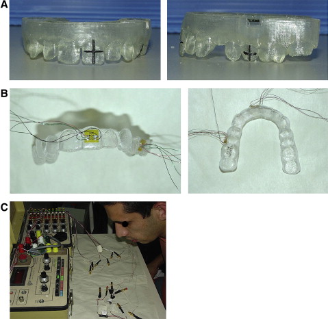
Maxillary and mandibular dental impressions were taken with polyvinyl siloxane used with 2-step putty (ESPE Express, 3M, St Paul, Minn) and wash (Coltene President, 3M) technique. Align Technology manufactured 2 identical series of maxillary aligners according to the treatment plan. Series 1 was worn by the patient during the biweekly course of treatment, and series 2 was used for the in-vivo von Mises strain measurements worn by the patient only during strain measurements ( Fig 1 ). Series 2 aligners received 2 strain gauge (SG) rosettes (EA-06-015RJ-120, Vishay Measurements Group, Raleigh, NC), 1 bonded on the aligner facing the labial aspect of the maxillary central incisor (active dental unit), and the other facing the buccal aspect of the maxillary first or second premolar (passive dental unit) that served as the anchorage tooth ( Fig 1 , B ). For accurate duplication of the SG position between aligners, a cross-bisection the midpoint of the crown was marked on in the SLA molds on the crown of the selected incisor and premolar ( Fig 1 , A ).
Measurements of series 2 were taken on day 1, when the new aligner of series 1 was given to the patient, and on days 2, 9, and 15. Day 15 was the day when the old aligner of series 1 was replaced by a new one (after 2 weeks) and was also day 1 for the first aligner of series 2.
Before each measurement, all 6 SGs (2 rosettes) of the series 2 aligners were connected in a quarter Wheatstone bridge via a switch and balance unit (SB-10, Vishay Measurements Group) to a strain indicator (P-3500, Vishay Measurements Group) ( Fig 1 , C ), and a zero extraoral baseline was established outside the mouth. Subsequently, the aligner was placed in the mouth, and intraoral strains were measured. All measurements were repeated 3 times. Differences between the extraoral and intraoral means of each of the 3 components of the SG rosette were calculated. From these values, principal strains were calculated and converted into a single value of von Mises strain. The von Mises strain is based on the distortion energy of a structure and is widely used in biomechanical studies. The incisor von Mises (IVM) strains and premolar von Mises (PVM) strains were obtained from the incisor and premolar rosettes, respectively.
Distributions of the calculated variables (von Mises strains) were analyzed with the Kolmogorov-Smirnoff test. Differences between aligners were analyzed with analysis of variance (ANOVA) with repeated measures with 2 factors: location (incisor and premolar) and time (days 1, 2, 9, and15). Measurements for variations were analyzed between aligners for each patient.
Results
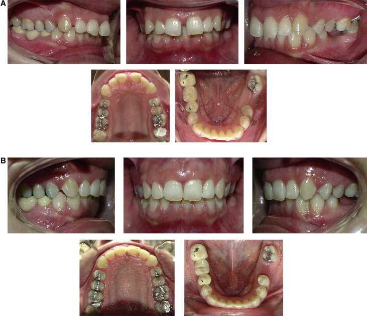
The treatment plan for patient 1 included closure of the diastema between the maxillary central incisors (9 aligners) and retraction of all 6 maxillary anterior teeth to reduce the overjet (29 aligners) ( Fig 2 ).
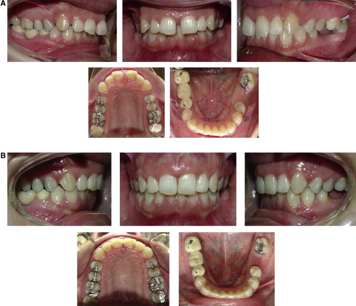
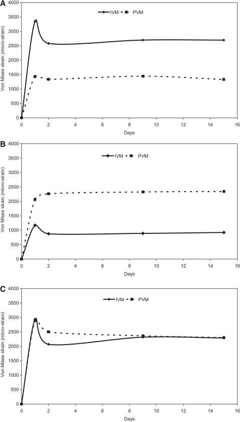
Figure 3 , A , and the Table give the mean values of IVM and PVM strains measured during maxillary incisor retraction treatment. Both variables (time and location) significantly affected the developed strains ( P <0.001). Additionally, an interaction was found between time and location ( P <0.005), indicating that time had a diverse influence on the strains measured in the 2 locations. A statistically significant difference was found between IVM strains obtained during 15 days of wearing each aligner ( P <0.001), with mean IVM strains measured on day 1 significantly higher (23%) than on day 2 and all other days ( P <0.001). No statistical differences were found between days 2, 9, and 15. However, for the premolar location, no significant difference was found between PVM strains during 15 days of wearing each aligner ( P = 0.626). When the day-1 peak IVM and the corresponding day-1 PVM between the sequential aligners were compared, it was found that, with an increase in the order of sequential aligners, day-1 peak IVM increased, and day-1 PVM decreased ( Fig 4 , A ). This same effect was found for day-15 von Mises strains ( Fig 5 , A ). Mean von Mises strains at all time points were about 2 times greater at the incisor location compared with the premolar.
| Day | Parameter | Mean | SE | Mean | SE | Mean | SE |
|---|---|---|---|---|---|---|---|
| Patient 1 | Patient 2 | Patient 3 | |||||
| 1 | IVM | 3344 a | 152 | 1174 a | 237 | 2931 a | 384 |
| PVM | 1431 c | 101 | 2068 c | 237 | 2889 a | 252 | |
| 2 | IVM | 2583 b | 170 | 877 b | 165 | 2071 b | 336 |
| PVM | 1336 c | 132 | 2264 c | 237 | 2500 a | 350 | |
| 9 | IVM | 2700 b | 150 | 891 a | 164 | 2316 b | 407 |
| PVM | 1445 c | 72 | 2327 c | 218 | 2363 b | 238 | |
| 15 | IVM | 2697 b | 168 | 923 a | 165 | 2288 b | 365 |
| PVM | 1332 c | 76 | 2345 d | 227 | 2300 b | 235 | |
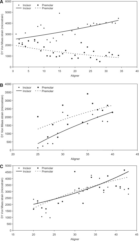
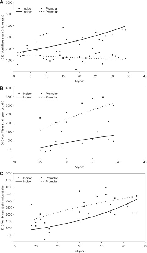
Measurements in patient 2 were taken during retraction of the maxillary central incisors from aligner number 35 up to aligner 47 (13 aligners). Figure 3 , B , and the Table show the mean values of IVM and PVM strains of the aligners measured during retraction. The location significantly affected the strains ( P <0.001), and an interaction was found between time and location ( P <0.006). However, for the premolar, no significant change was found regarding time ( P >0.546). Mean IVM strains measured on day 1 were significantly higher (43%) than those measured on day 2 ( P <0.03), but differences between days 1 and 9 ( P = 0.062) or day 15 ( P = 0.081) were nonsignificant, with day 1 having higher strain values (40%). In contrast, mean PVM strains measured on day 1 were nonsignificantly lower (9%) compared with days 2 and 9 ( P = 0.074, P = 0.073, respectively) and significantly lower compared with day 15 ( P <0.038). When the day-1 peak IVM and the corresponding day-1 PVM between the sequential aligners were compared, it was found that, with an increase in the order of sequential aligners, both day-1 peak IVM and day-1 PVM increased; however, day-1 peak IVM was lower than day-1 PVM ( Fig 4 , B ). This same effect was found for day 15 von Mises strains ( Fig 5 , B ).
For patient 3, Von Mises measurements were collected from 19 aligners during incisor retraction. Figure 3 , C , and the Table present the mean values of IVM and PVM of all aligners measured during treatment. In both locations, time similarly influenced the developed von Mises strains ( P <0.001). However, the location did not affect the von Mises strains ( P = 0.832). No interaction was found between time and location ( P = 0.299), indicating that time had a similar effect on the von Mises strains measured in the 2 locations. A statistically significant difference was found between IVM strains obtained during the 15 days of wearing each aligner ( P <0.001), with significantly greater strains measured on day 1 (29%) than on day 2 and all other days ( P <0.001) and no significant differences between the other days. However, no significant difference between PVM strains was found between days 1 and 2, but day 1 had significantly higher values than days 9 and 15 ( P <0.001). When the day-1 peak IVM and the corresponding day-1 PVM between the sequential aligners were compared, it was found that, with increase in the order of sequential aligners, both day-1 peak IVM and day-1 PVM increased, and the day-1 peak IVM was slightly higher than day-1 PVM ( Fig 4 , C ). This same effect was found for the day-15 von Mises strains ( Fig 5 , C ).
Results
The treatment plan for patient 1 included closure of the diastema between the maxillary central incisors (9 aligners) and retraction of all 6 maxillary anterior teeth to reduce the overjet (29 aligners) ( Fig 2 ).

