Introduction
The objective of this study was to evaluate the surface enamel after bracket debonding and residual resin removal.
Methods
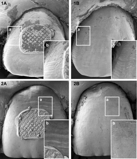
Thirty patients (female, 20; male, 10; mean age, 18.4 years) who completed orthodontic treatment with fixed appliances (Twin Brackets, 3M Unitek, Monrovia, Calif) (n = 525) were included. The amounts of adhesive left on the tooth surfaces and the bracket bases were evaluated with the adhesive remnant index (ARI). ARI tooth (n = 498) was assessed on digital photographs by 2 operators. After resin removal and polishing, epoxy replicas were made from the maxillary anterior teeth (n = 62), and enamel surfaces were scored again with the enamel surface index. Elemental analysis was performed on the debonded bracket bases by using energy dispersive x-ray spectrometry mean area scanning analysis. The percentages of calcium and silicon were summed up to 100%. Tooth damage was estimated based on the incidence of calcium from enamel in relation to silicon from adhesive (Ca%) and the correlation between the ARI bracket and Ca%.
Results and Conclusions
While ARI tooth results showed score 3 as the most frequent (41%) ( P <0.05), followed by scores 0, 1, and 2 (28.7%, 17.9%, and 12.4%, respectively), ARI bracket results showed score 0 more often (40.6%) than the other scores ( P <0.05). Maxillary anterior teeth had significantly more scores of 3 (49%) than the other groups of teeth (10%-25%) (chi-square; P <0.001). There were no enamel surface index scores of 0, 3, or 4. No correlation between the enamel surface index and ARI tooth scores was found (Spearman rho = 0.014, P = 0.91). The incidence of Ca% from the scanned brackets showed significant differences between the maxillary and mandibular teeth (14% ± 8.7% and 11.2% ± 6.5%, respectively; P <0.05), especially for the canines and second premolars (Kruskal-Wallis test, P <0.01). With more remnants on the bracket base, the Ca% was higher (Jonckheere Terpstra test, P <0.05). Iatrogenic damage to the enamel surface after bracket debonding was inevitable. Whether elemental loss from enamel has clinical significance is yet to be determined in a long-term clinical follow-up of the studied patient population.
Editor’s comment
Structural and colorimetric alterations after debonding have been studied by a number of in-vitro and in-vivo protocols in the past. Surface roughness, enamel removal, and color change have been investigated with various techniques. This study is one of the few available that reports data from in-vivo conditions. The authors sought to evaluate the surface enamel after bracket debonding and residual resin removal. Thirty patients who completed orthodontic treatment with fixed appliances were included. Two operators examined digital photographs and applied the adhesive remnant index (ARI) to rate the amount of adhesive left on the tooth surface (ARI tooth ); adhesive remaining on the bracket base (ARI bracket ) was also evaluated. After resin removal and polishing, epoxy replicas were made from the maxillary anterior teeth, and enamel surfaces were scored again with the enamel surface index. Elemental analysis was performed on the debonded bracket bases by using energy dispersive x-ray spectrometry mean area scanning analysis. The percentages of calcium and silicon were summed up to 100%. Tooth damage was estimated based on the incidence of calcium from enamel in relation to silicon from adhesive (Ca%) and the correlation between the ARI bracket and Ca%. This investigation showed that ARI tooth showed score 3 as the most frequent (41%), followed by scores 0, 1, and 2 (28.7%, 17.9%, and 12.4%, respectively). ARI bracket results showed score 0 more often (40.6%) than the other scores. Maxillary anterior teeth had significantly more scores of 3 (49%) than other groups of teeth (10%-25%). There were no enamel surface index scores of 0, 3, and 4. No correlation between the enamel surface index and ARI tooth scores was found. The incidence of Ca% from scanned brackets showed significant differences between the maxillary and the mandibular teeth (14% ± 8.7% and 11.2% ± 6.5%, respectively), especially for canines and second premolars. The more adhesive remnants on the bracket base, the higher the Ca%. Iatrogenic damage to the enamel surface after bracket debonding was inevitable.


