(1)
Department of Pathology, Radboud University Nijmegen Medical Center, Nijmegen, The Netherlands
Abstract
As already mentioned before, immature odontogenic tissues may closely mimic odontogenic tumours. Quite often, it may be very difficult to decide if the structures visible in the slide are indicating the presence of an odontogenic tumour or that they are just remnants of tissues connected with tooth development. Two different parts of the immature tooth are important in this respect: the dental papilla and the dental follicle. Both will be discussed in some detail.
Introduction
As already mentioned before, immature odontogenic tissues may closely mimic odontogenic tumours. Quite often, it may be very difficult to decide if the structures visible in the slide are indicating the presence of an odontogenic tumour or that they are just remnants of tissues connected with tooth development [1–3]. Two different parts of the immature tooth are important in this respect: the dental papilla and the dental follicle. Both will be discussed in some detail.
Before, it has to be emphasised that one should be very cautious in making a diagnosis of some kind of odontogenic tumour on tissue taken from a jaw area still containing developing teeth and not showing any clinical or radiographic sign of tumour being present. The ubiquitousness of immature odontogenic tissues in the jaw of children is illustrated in Fig. 12.1. This picture makes understandable that the chance of encountering immature odontogenic tissues in tissue samples from this area and at this age is rather high.
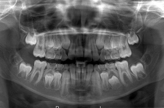
Fig. 12.1
Radiograph showing mixed dentition. Germs of the permanent teeth in varying stages of maturation are seen in upper and lower jaw. The third molar germ has just started to form dental hard tissues, whereas the second permanent molar germ shows a very pronounced radiolucent rim representing the dental follicle. Other tooth germs show wide apical foramina indicating incompletely formed roots. Histologic details from this root area are shown in Fig. 12.2 (Picture kindly provided by Prof. A. Kuijpers-Jagtman, Department of Orthodontics and Craniofacial Biology, Radboud University Nijmegen Medical Center, Nijmegen, The Netherlands)
Dental Papilla
The dental papilla is the mesenchymal soft tissue part of the tooth germ. During development and maturation, it gradually transforms into the dental pulp. Concomitant with this transformation, it decreases in volume and changes its histomorphology from mainly myxoid to more fibrous. Due to this myxoid nature, the dental papilla can be mistaken for odontogenic myxoma with serious consequences for the patient [1–3].
This diagnostic pitfall can be avoided by an enquiry into the source of the tissue. If there is evidence that it comes from the vicinity of a developing tooth, dental papilla is much more likely to be the appropriate diagnosis than odontogenic myxoma. Moreover, in case of dental papilla, the myxoid tissue quite often is accompanied by some strands of odontogenic epithelium representing remnants of the enamel organ, and also, it may show a peripheral cell condensation indicating presence of odontoblasts. Finally, a shell of dentin adjacent to the myxoid tissue may be helpful in differentiating odontogenic myxoma from dental papilla (Figs. 12.2 and 12.3a–d).
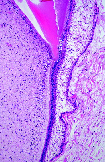
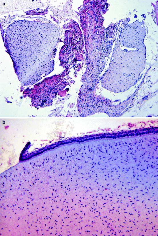
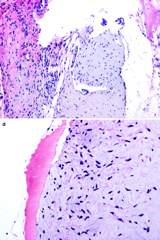
Fig. 12.2
Photomicrograph of the apical part of a tooth germ. Dental papilla (left side) consisting of immature myxoid connective tissue faces dentin and inner enamel epithelium (right side). Interposed cell condensation represents layer of odontoblasts forming dentin. See also figures in Chap. 1
Fig. 12.3
Dental papilla mimicking myxoma. (a) Low-power view showing fragments of loose myxoid tissue are surrounded by more dense fibrous bands. (b) Adjacent strand of inner enamel epithelium reveals that it concerns a dental papilla and not a myxoma. (c) Sometimes, the epithelial component is only rudimentary. (d) Adjacent tubular dentin also indicates that it does not concern a myxoma but normal immature odontogenic tissue
In practice, this diagnostic pitfall especially applies to biopsies taken from elevations of the bottom of the maxillary sinus. These elevations may well represent the cranial part of developing maxillary tooth germs. If biopsied and submitted as tissue from a swelling in the maxillary sinus, an erroneous diagnosis of maxillary odontogenic myxoma is easily made [4].
Dental Follicle
The dental follicle is the fibrous sac that surrounds the not-yet-erupted immature tooth (Fig. 12.4a, b). At radiographs, it can be seen as a radiolucent rim surrounding the tooth germ (Fig. 12.5). Sometimes, this rim is larger than usually seen. This may lead to further examination including biopsy. As these dental follicles may consist of myxoid tissue, they may be mistaken for myxoma similar as for the dental pulp [2, 5
Stay updated, free dental videos. Join our Telegram channel
VIDEdental - Online dental courses


