(1)
Department of Pathology, Radboud University Nijmegen Medical Center, Nijmegen, The Netherlands
Abstract
Teeth consist for the major part of dentin. This material houses the dental pulp, the soft tissue core of the tooth consisting of myxoid connective tissue with blood vessels and nerves, and supports the enamel cap that covers the part of the tooth that is exposed to the oral cavity. In the root area, dentin is covered by cementum that fixes the collagenous fibres of the periodontal ligament onto the root surface. At the other side, these collagenous fibres are attached to the bone of the tooth socket, and in this way, the tooth is fixed in the jaw. Through an opening at the root tip that is called the apical foramen, the connective tissue of the pulp is continuous with the collagenous fibres of the periodontal ligament. Blood vessels and nerves pass through this opening to the dental pulp (Fig. 2.1a, b). Sometimes, additional communications exist between dental pulp and periodontal ligament. These so-called accessory canals are clinically important as they may cause lesions usually confined to the root tip to occur at aberrant sites.
Introduction
Teeth consist for the major part of dentin. This material houses the dental pulp, the soft tissue core of the tooth consisting of myxoid connective tissue with blood vessels and nerves, and supports the enamel cap that covers the part of the tooth that is exposed to the oral cavity. In the root area, dentin is covered by cementum that fixes the collagenous fibres of the periodontal ligament onto the root surface. At the other side, these collagenous fibres are attached to the bone of the tooth socket, and in this way, the tooth is fixed in the jaw. Through an opening at the root tip that is called the apical foramen, the connective tissue of the pulp is continuous with the collagenous fibres of the periodontal ligament. Blood vessels and nerves pass through this opening to the dental pulp (Fig. 2.1a, b). Sometimes, additional communications exist between dental pulp and periodontal ligament. These so-called accessory canals are clinically important as they may cause lesions usually confined to the root tip to occur at aberrant sites.
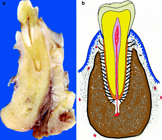
Fig. 2.1
(a) Gross specimen showing tooth in its socket with surrounding jaw bone. (b) schematic drawing to clarify the various components: enamel in grey, dentin in yellow, root cementum in orange, periodontal ligament in green and epithelium in blue (Modified from drawing by John A.M. de Groot)
Dentin
Dentin is a specialised kind of bone formed by the odontoblasts but different in that sense that it does not contain complete cells but only cellular extensions, cytoplasmic extensions from the odontoblasts. These cross the full thickness of the dentin from the odontoblastic cell body that lies at the border between dentin and dental pulp to the junction between dentin and enamel. The tiny canals that house the odontoblastic extensions are recognisable as evenly spaced tubuli. This tubular nature is the histologic hallmark for dentin, not only in teeth but also in odontogenic lesions in which the nature of each mineralised material may not be recognisable at first sight. Sometimes, in the dentin incremental lines that run parallel to the dentino-enamel junction, lines are observed: the lines van von Ebner.
The organic matrix laid down by the odontoblasts is called predentin recognisable by its pink staining in hematoxylin and eosin-stained sections. In this matrix, calcification occurs in the form of spherical masses which, as they increase in size, gradually fuse together, thus transforming the predentin into the darker-staining mineralised dentin (Fig. 2.2). At the border between dentin and predentin, these calcospherites can be frequently seen, either separate or already in contact with the calcified dentin.
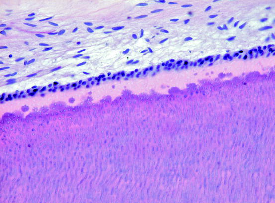
Fig. 2.2
Dentin at its pulpal side bordered by columnar odontoblasts. The distinction between the pale predentin and the darker-staining mineralised dentin is clearly visible
Several types of dentin are discerned. The layer of dentin immediately adjacent to the border with the enamel is called the mantle dentin. The more centrally located part of the dentine is the circumpulpal dentin. This distinction is relevant as some structural tooth alterations are confined to the circumpulpal dentin, leaving the mantle dentine uninvolved as will be discussed later on. Mantle dentin is separated from the circumpulpal dentin by a zone of disturbed dentin formation called globular dentin, named in this way because of the presence of interglobular spaces due to deficient mineralisation at this site (Fig. 2.3
Stay updated, free dental videos. Join our Telegram channel
VIDEdental - Online dental courses


