and Jasdeep Kaur1
(1)
Earth and Life Sciences Vrije Universiteit Amsterdam and ILEWG, Amsterdam, The Netherlands
11.1 Introduction
11.2.1 Periapical Radiographs
11.2.2 Bitewing Radiographs
11.2.3 Occlusal Radiographs
11.3 Extraoral Techniques
11.3.2 Lateral Oblique Jaw View
11.3.3 Cephalometric View
11.3.4 Temporomandibular Joint View
11.4 NOMAD
11.5.1 Age Estimation
11.5.2 Sex Determination
11.5.3 Racial Determination
11.10 Conclusions
Abstract
Orodental radiographs are a very important tool in identification in forensic odontology. Different methods for the identification of human remains exist, but most are based on comparisons between available ante- and postmortem data. Also, forensic dental radiography helps in the construction of a profile when antemortem records are not available. This chapter discusses the role of orodental radiography in forensic odontology.
11.1 Introduction
Recently, forensic radiology has been based almost exclusively on X-rays and images captured on the radiograph. The advanced modalities, such as computed tomography (CT), cone beam computed tomography (CBCT), and magnetic resonance imaging (MRI), are being added to the forensic toolkit. The X-ray fingerprint method known as X-ray fluorescence radiography (XFR) has been revived in the recovery of latent prints from surfaces. The XFR method is employed when prints are suspected to rest on problematical materials such as multicolored documents, cloth, polythene, wax, cardboard, hardboard, varnished and untreated wood, rubber pigskin, and the skin of human corpses or nonvital separated body parts. This method is not used commonly because it needs special tools, and the lead dust can expose forensic investigators to a health risk (American College of Radiology 2011).
The forensic odontologist identifies and characterizes the teeth of unidentified bodies and essentially works expansively with radiographs. The dental charts of missing individuals can then be compared with the forensic odontologist’s report to identify the body. If the body is decomposed, skeletonized, fragmented, burned, or mutilated for any other reason, it is extremely common that the dentition will be intact and will provide a valuable tool for the identification process (Kvaal et al. 1995; Lichtenstein et al. 1988). This is predominantly true for victims of fires and mass disasters (Ligthelm 1983). Thus, despite the abundance of possible techniques, those used in forensic dentistry are extremely valuable for this purpose (Sainio et al. 1990). Thus, dental records and radiographs are the most useful tool for identifying such mutilated remains (Fischman 1985). This chapter will highlight the scope of oral-dental radiology in forensic odontology.
11.2 Intraoral Radiographic Techniques
Two of the basic principles of radiography are that (1) the central beam must pass through the area to be examined, and (2) the X-ray film must be placed in position so as to record the image with the least amount of image distortion (Dunn and Kantor 1993).
11.2.1 Periapical Radiographs
The principle of the intraoral periapical (Fig. 11.1) examination is to acquire a view of the tooth and its surrounding structures. Two exposure techniques may be employed for periapical radiography: the paralleling technique and the bisecting angle technique. The paralleling technique is the most common method. This technique gives less image distortion and reduces excess radiation exposure to the patient. The bisecting technique can be applied for subjects unable to accommodate the positioning of the paralleling technique. Shortcomings to the bisecting technique comprise image distortion and excess radiation due to increased angulations involving the eye and thyroid glands. The paralleling technique is so named because the tooth, receptor, and end of the position-indicating device are all maintained on parallel planes. The principle is based on the fact that image sharpness is primarily affected by the focal-film distance, object film distance, motion, and the effective size of the focal spot of the X-ray tube. It requires the use of film holding devices to maintain the relatively particular positioning needed. A variety of film holders are available: simple, complex, light, heavy, reusable, disposable, autoclavable, and non-autoclavable. A few of the more common include XCP with localizing rings, Snap-a-ray, and Stabe Bite Blocks. In the bisecting technique, the film touches the teeth at the incisal edge or the lingual-occlusal surface and then the rest of the film is angled depending on the anatomy of the area. The X-ray beam is directed perpendicular to the bisecting line between the angle of the long axis of the tooth and the angle of the film. Supporting the film pack with the patient’s forefinger is not recommended.
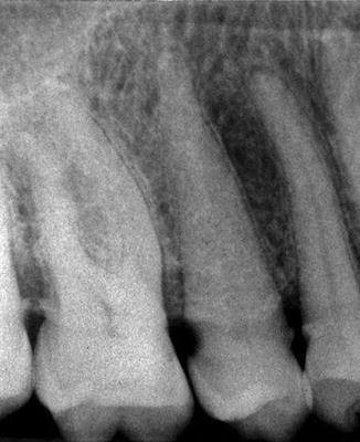
Fig. 11.1
Periapical radiograph
11.2.2 Bitewing Radiographs
Bitewing X-rays (Fig. 11.2) show the size of the pulp chamber. They are a useful adjunct to evaluate periodontal conditions, offer a good view of the septal alveolar crest, and, in addition, permit changes in bone height to be accurately assessed by comparison with adjacent teeth. They do not provide information about the apices of the tooth.
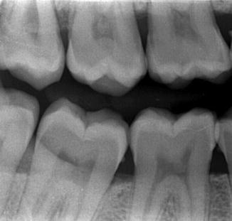
Fig. 11.2
Bitewing radiograph
11.2.3 Occlusal Radiographs
Occlusal radiographs (Fig. 11.3) allow a complementary radiographic examination intended to provide a more extensive view of the maxilla and mandible. They are very useful in determining the buccolingual extension and displacement of fractures of the mandible and maxilla. They also aid in localizing unerupted teeth, retained roots, foreign bodies, and calculi in the submandibular and sublingual salivary glands and ducts.
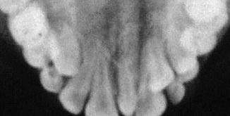
Fig. 11.3
Occlusal radiograph
11.3 Extraoral Techniques
Various extraoral techniques have been proposed, which this section reviews (Fischman 1985).
11.3.1 Panoramic Radiograph (Hatcher 2010)
A panoramic X-ray (Fig. 11.4) shows the dentition and surrounding structures, the facial bones and condyles, and parts of the maxillary sinus and nasal complexes. The panoramic radiography equipment comprises a horizontal rotating arm that holds an X-ray source and a moving film mechanism (carrying a film) arranged at opposed extremities. The subject’s head sits between the X-ray generator and the film. The X-ray source is collimated toward the film, to provide a beam shaped as a vertical blade having a width of 4–7 mm (Hatcher 2010
Stay updated, free dental videos. Join our Telegram channel
VIDEdental - Online dental courses


