Introduction
The aim of this study was to apply Bayesian networks to evaluate the relative role and possible causal relationships among various factors affecting the diagnosis and final treatment outcome of impacted maxillary canines.
Methods
A total of 168 patients with infraosseous impacted maxillary canines had a combined surgical-orthodontic approach aimed to guide the impacted tooth to the center of the alveolar ridge. Demographic, orthodontic, and periodontal variables were recorded and analyzed by means of Bayesian network analysis.
Results
All 168 impacted canines were successfully moved and aligned in the dental arches with healthy periodontiums. According to the Bayesian network analysis, bilateral impaction was associated with palatal impaction and longer treatment; the pretreatment α-angle was a determinant for the duration of orthodontic traction, also because of the associations between greater angulation of impacted canines with more severe tooth displacement and with greater distance of the impacted canine from the occlusal plane; the posttreatment periodontal outcome was not related to the pretreatment radiographic variables.
Conclusions
Bayesian network analysis was useful to identify possible relationships among the variables considered for diagnosis and treatment of impacted canines.
The prevalence of impacted canines was reported to be from 0.2% to 2.8% according to several authors. An impacted canine requires a complex therapeutic management, which can be considered successful only if the forced eruption and the subsequent alignment lead the tooth to the correct position in the dental arch with a healthy periodontium. The eruption of the tooth between the alveolar cortical plates prevents bone dehiscence and unfavorable orthodontic and esthetic consequences. Therefore, the most appropriate treatment should simulate the physiologic eruption pattern that occurs at the center of the alveolar ridge, as some authors have suggested. Orthodontic appliances and techniques specifically designed for this purpose have been proposed. In case of persistent deciduous canines and unerupted permanent canines, the “tunnel” technique might be indicated to reproduce the physiologic eruption pattern of the canine.
The therapeutic approach to impacted canines is interdisciplinary, with many factors accounting for the final orthodontic and periodontal outcomes. Pretreatment radiographic features of impacted canines—α-angle, d-distance, and sector of impaction according to Ericson and Kurol —have been shown to be predictive factors for the durations of orthodontic traction and comprehensive orthodontic treatment to reposition the impacted tooth. The more severely displaced the canine with regard to the adjacent maxillary incisors, the longer the orthodontic treatment. The indicators on pretreatment panoramic films have been studied also as predictors for the outcomes of interceptive treatment of palatally displaced canines by means of extraction of the corresponding deciduous canine and space maintenance in the maxillary dental arch. However, the same radiographic variables had no predictive value for the final periodontal status of the impacted canines after surgical-orthodontic treatment to reposition the canine at the center of the alveolar ridge. Most investigations evaluated the relationships between factors accounting for treatment outcomes of impacted canines with descriptive statistics or linear regression on a priori identified variables; more recent studies used multilevel statistics to study associations among factors without determining causal relationships.
Bayesian networks (BN) were introduced recently with the goals of generating hypotheses of possible causal relationships among variables and promoting further specific studies (ie, randomized clinical trials). BN adopt an intermediate approach between statistics and artificial intelligence. A “network” is composed of a “directed acyclic graph” in which stochastic variables are represented by vertices or nodes of the graph, and oriented lines (arrows) represent the relationships among the variables. The arrows relate the variables in such a way that cycles are not permitted, so that, following the arrows, it is impossible to return to a vertex or starting point. The variables from which the arrows start influence those to which they arrive, possibly through a causal relationship. An example of Bayesian analysis was reported in an oral oncology genomic study, and some aspects of a directed acyclic graph have been elucidated in dental research. Recently, BN have been applied to the analysis of relevant literature in implantology. At the present time, no study applies BN analysis in orthodontics.
The aim of this investigation was to apply BN to comprehensive surgical-orthodontic treatment of maxillary impacted canines to evaluate the relative role and the possible causal relationships among various factors affecting the clinical approach to this condition.
Material and methods
Our subjects were 168 patients with unilateral or bilateral infraosseous impacted maxillary canines from a previous study. One hundred twenty-five patients had unilateral impaction of the maxillary canine, and 43 had bilateral impactions. A random selection was made of the 86 bilateral impacted canine to evaluate only 1 canine per patient. The final study population consisted of 168 patients (168 impacted canines) (40 male, 128 female; age range, 12.8-52.0 years; mean age, 17.2 ± 6.0 years).
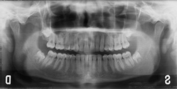
The following variables before treatment were collected: (1) buccal or palatal site of impaction, left or right side, unilateral or bilateral impaction; (2) radiographic variables on panoramic x-rays: α-angle: angle measured between the long axis of the impacted canine and the midline, d-distance: distance between the canine cusp tip and the occlusal plane (from the first molar to the incisal edge of the central incisor), and s-sector: sector where the cusp of the impacted canine is located (sector 1, between the midline and the axis of the central incisor; sector 2, between the axes of the central incisor and the lateral incisor; or sector 3, between the axes of the lateral incisor and the first premolar).
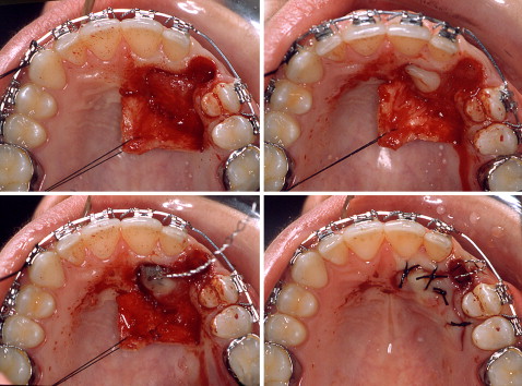
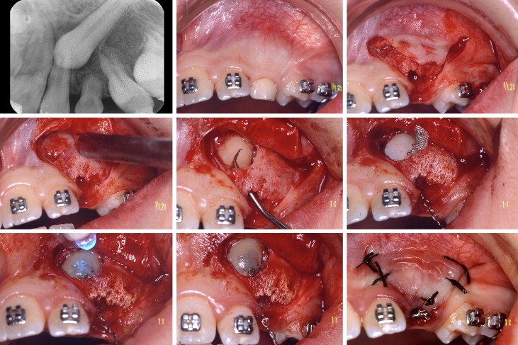
All patients underwent consecutively a closed-flap surgical approach followed by orthodontic alignment. Treatment was delivered by 1 operator (A.C.) in a time span of 17 years. The teeth were exposed by means of a repositioned flap. Orthodontic traction was applied to guide the impacted canine directly toward the center of the alveolar ridge. In patients with persistent deciduous canines and unerupted permanent canines (n = 24), the “tunnel” technique was used.
The overall combined treatment was divided into 3 phases.
- 1.
Initial orthodontic treatment was aimed at creating space in the maxillary arch with fixed appliance therapy.
- 2.
Surgical exposure and orthodontic traction were used to move the impacted tooth toward the center of the alveolar ridge. A handmade chain was connected to the attaching device on the impacted tooth and to the elastic for orthodontic traction. A rectangular stabilization arch was used to obtain adequate anchorage and maintain sufficient space in the dental arch, and a round arch was used as an attachment for the elastic traction to guide the impacted canine toward the center of the alveolar ridge. The duration of this phase (duration of traction) was calculated as the time between the application of the traction device and the emergence of the cusp of the impacted canine.
- 3.
Final orthodontic treatment aligned the canine in the maxillary arch.
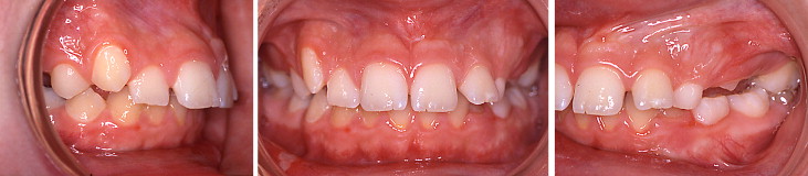
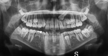
Two patients are shown in Figures 1 to 10 .


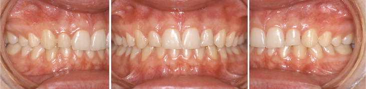
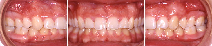
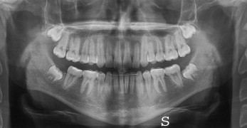
The treated teeth were evaluated periodontally after the overall orthodontic treatment (phases 1-3).
The following periodontal variables were considered for the treated canines.
- 1.
Probing depth (PD) measurements were made with a Williams offset periodontal probe at 6 sites—mesiobuccal, midbuccal, distobuccal, mesiolingual, midlingual, and distolingual—of each treated tooth. The greatest PD was used in the analysis.
- 2.
Width of keratinized tissue (KT), from the gingival margin to the mucogingival junction, was measured at the medial position of the buccal aspect of the crown.
Statistical analysis
Descriptive statistics were calculated as means and standard deviations for metric variables and as frequencies for nominal variables. An automatic structural learning algorithm of the BN was used as an explorative statistical technique for detecting possible causal relationships among these variables: (1) demographic variables (sex and age); (2) topographic variables (clinical and radiographic): site (buccal or palatal), side (left or right), unilateral or bilateral (patient), α-angle, d-distance, s-sector; treatment technique (tunnel); duration of traction, duration of treatment; KT; and PD.
The metric variables were transformed into binary variables by using the median values as a threshold. For the variable age, the threshold of 20 years was used. The variable s-sector was transformed into a binomial variable by combining sectors 1 and 2. For the generation of the directed acyclic graph, the structural learning algorithm B was used, and the variables were organized in 5 levels. These levels (temporal tiers [TT]) imply a hierarchic order, so that subsequent levels cannot influence previous ones: TT1, sex and age; TT2, site (buccal or palatal), side (left or right), unilateral or bilateral (patient), α-angle, d-distance, and s-sector; TT3, treatment technique (tunnel); TT4, duration of traction; and TT5, duration of treatment, KT, and PD.
To exemplify the concept of the TT, the amount of KT after therapy cannot influence the side of canine impaction. By using these limitations, the graph that illustrates the relationships among the variables was generated.
Stay updated, free dental videos. Join our Telegram channel
VIDEdental - Online dental courses


