Introduction
The smile is a key facial expression, and a careful assessment of the facial profile in smiling is an essential part of a complete orthodontic diagnosis. The aim of this study was to determine the preferred maxillary incisor inclination in the smile profile with regard to different mandibular positions.
Methods
A smiling profile photograph of a man with normal facial profile features was altered digitally to obtain 3 different mandibular sagittal positions in 4-mm decrements or increments from −4 to +4 mm. In each mandibular position, the inclination of the maxillary incisors was changed from −10° to +10° in 5° increments. A total of 234 raters (72 senior dental students, 24 orthodontists, 21 maxillofacial surgeons, 25 prosthodontists, and 92 laypeople) were asked to score each photograph using a Likert-type rating scale. Mann-Whitney, Kruskal-Wallis, and intraclass correlation coefficient tests were used to analyze the data.
Results
In retruded and protruded mandibles, normal incisor inclination and the most retroclined incisors were selected as the most and the least attractive images, respectively, by almost all groups. With an orthognathic mandible, the image with the most retroclined incisors was selected as the least attractive, but the raters were not unanimous regarding the most attractive image. The intraclass correlation coefficient was 0.82 (high level of agreement). Also, the sex of the raters had no effect on the rating of the photographs.
Conclusions
It is crucial to establish a normal incisor inclination, especially in patients with a mandibular deficiency or excess. An excessive maxillary incisor lingual inclination should be avoided regardless of the mandibular position.
Highlights
- •
Normal maxillary incisor inclination is favored when the mandible is retruded.
- •
Normal maxillary incisor inclination is favored when the mandible is protruded.
- •
When the mandible is positioned normally, the preferred incisal inclination varies.
- •
Raters generally prefer less incisor inclination.
- •
Raters prefer labial inclination over lingual inclination.
Improving facial esthetics has gained more popularity with the advent of the soft tissue paradigm and is a main goal in the treatment of orthodontic patients. The mouth is an important feature in facial attractiveness, and a facial smiling profile assessment is an integral part of a complete orthodontic diagnosis. Kerns et al reported that from an esthetic viewpoint, the profile and frontal views of the same smile were not rated similarly; the former was rated higher than the latter. Buccolingual inclination of the maxillary incisors also plays a major role in profile smile attractiveness.
To improve the prediction of the most proper position of the maxillary incisors, several profilometric studies have been conducted. Schlosser et al compared the preferences of orthodontists and laypeople with regard to the buccolingual position of the maxillary incisors in smiling profiles. This study showed a higher level of acceptance with maxillary incisor protrusion than with retrusion in both panels and therefore suggested either not to retract a normally protrusive maxillary dentition or to advance rather than retract the maxillary anterior teeth. In another study by Ghaleb et al, 3 groups including dentists, orthodontists, and laypeople scored the attractiveness of smiling profiles based on maxillary incisor inclinations. The results showed that a 5° protrusion of the maxillary incisors from the normal inclination had the highest rate of appeal among the raters. A statistically significant difference was found among different groups regardless of the sex of the raters of the preferred profile photographs.
Cao et al reported that the smiling profile with a 5° lingual incisor inclination was the most favorable among their panels (orthodontists and undergraduate students), whereas the profiles with 15° of labial inclination received the lowest scores.
Although previous studies of profile esthetics have mainly focused on the position or the inclination of the maxillary incisors in profile views with normal mandibular position, to our knowledge, no studies have yet evaluated the esthetic effects of maxillary incisor inclination with regard to different mandibular positions in smiling profiles. Moreover, the mandibular position is a characteristic of the patient’s inherent underlying skeletal pattern and is difficult to alter during orthodontic treatment. Therefore, it may be important for clinicians to take into account the balance between the incisor inclination and the mandibular sagittal position. This information might assist orthodontists in considering mandibular position in treatment planning for choosing the appropriate inclination for the maxillary incisors.
The objectives of this study were to determine the preferred maxillary incisor inclination in the smile profile of a male subject with regard to different mandibular positions and to elucidate whether the raters’ profession and sex played a role in the assessment of the preferred maxillary incisor inclination.
Material and methods
A finished orthodontic patient (age, 23 years) was chosen from the patients treated at the orthodontics clinic of Shiraz University of Medical Sciences. Informed consent was obtained from the patient for participating in this study. He was chosen based on the following clinical and lateral cephalometric criteria: (1) Class I canine and molar relationships with adequate overjet and overbite, (2) well-positioned maxillary incisors according to cephalometric standards, (3) normal facial convexity angle and vertical height ratio as described by Legan and Burstone, (4) normal soft tissue cephalometric analysis (Ricketts’ E-line and Merrifield’s z-angle ), and (5) facial angle and H-angle as described by Holdaway and nasolabial angle and maxillary lip angle as described by Arnett and Bergman within the normal range.
A right lateral profile photograph with the patient in natural head position with a blue background at a distance of 1.5 m from the camera and a speed of 1/125 was taken with a digital camera (c-2000; Olympus America, Melville, NY) under standard conditions. To standardize the photograph, the subject was asked to sit down. By using the ear positioners of the cephalostat, both the Frankfort plane and the pupillary plane were parallel to the ground.
The first image was taken with a neutral facial expression. The second image was taken with the subject in a posed smile, and a small 100-mm ruler was fixed above his head on the facial sagittal plane.
This 100-mm fixed ruler was used as a guide for computer-aided alterations to quantify hard and soft tissue alterations. The ruler and ear positioners of the cephalostat were later removed digitally to give the subject a normal appearance. The use of image alterations of 1 subject has been shown to be successful in studying variations in dental appearance.
The smiling photograph was altered using a commercially available image editing software program (Adobe Photoshop CS, version 8.0; Adobe Systems, San Jose, Calif). During the first alteration step, only 1 parameter was changed: the anteroposterior position of the mandible. The mandibular prominence of the subject’s facial profile was altered in 4-mm decrements and increments from −4 to +4 mm in to represent retrusion and protrusion of the mandible, respectively. By changing the position of the mandible in the horizontal plane relative to the true vertical line that crosses the glabella (defined as the most prominent anterior point in the midsagittal plane of the forehead ), 3 profiles were created (retruded, normal, and protruded). To focus on the sagittal aspect of the facial profile, the vertical height of the constructed face was kept constant.
In the next step, each profile group was further divided into 5 subgroups. The maxillary incisor inclination of each image was changed from −10° to +10° relative to the norm values of the subject in 5° decrements and increments to represent retroclined and proclined incisors. To simulate the changes of incisor inclination, the crowns of the central and lateral incisors were separately cut in the Adobe Photoshop program. Each tooth was considered as an individual object with the center of rotation at the incisal edge. The central incisor was superimposed from the tracing of the lateral cephalograms, and the center of rotation was placed at the incisal edge of the tooth. To maintain the symmetry, the center of rotation of the lateral incisor was set at the midpoint of the mesiodistal width. To maintain the vertical positions of the maxillary incisors, horizontal lines were drawn tangent to the incisal edges of the teeth, and vertical tangents were drawn medial to the maxillary canines as the distal limit for sagittal repositioning of the lateral incisor.
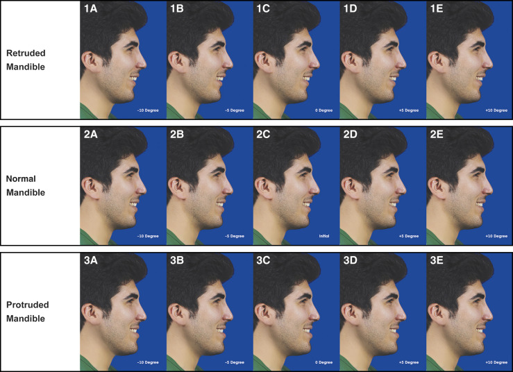
Each simulation was made in 5° decrements and increments, and 2 modifications were produced to represent retroclined incisors and 2 to represent proclined incisors. Artistic editing was used when necessary to maintain a natural appearance. Thus, overall, 3 sets of images were reproduced with different mandibular positions, and each set comprised 5 different maxillary incisor inclinations (from most retroclined to most proclined positions) ( Fig 1 , Table I ).
| Image | Situation |
|---|---|
| 1A | Retruded mandible with −10° of palatal incisor inclination |
| 1B | Retruded mandible with −5° of palatal incisor inclination |
| 1C | Retruded mandible with normal incisor inclination |
| 1D | Retruded mandible with +5° of labial incisor inclination |
| 1E | Retruded mandible with +10° of labial incisor inclination |
| 2A | Normal mandible with −10° of palatal incisor inclination |
| 2B | Normal mandible with −5° of palatal incisor inclination |
| 2C | Normal mandible with normal incisor inclination |
| 2D | Normal mandible with +5° of labial incisor inclination |
| 2E | Normal mandible with +10° of labial incisor inclination |
| 3A | Protruded mandible with −10° of palatal incisor inclination |
| 3B | Protruded mandible with −5° of palatal incisor inclination |
| 3C | Protruded mandible with normal incisor inclination |
| 3D | Protruded mandible with +5° of labial incisor inclination |
| 3E | Protruded mandible with +10° of labial incisor inclination |
Each series of images was printed separately on Digital Royal Paper (Kodak; Rochester, NY) with a Hewlett-Packard Photo Smart printer (Hewlett-Packard, Palo Alto, Calif) in a 15 × 20 cm format and then was placed randomly in a binder. The images were created so that each profile photograph had the same dimensions as a normal human head, based on an average lower anterior facial height. This helped to reduce the potential bias caused by image magnification or size reduction in the observer’s perception.
The rating panel comprised 234 raters including 24 orthodontists, 21 maxillofacial surgeons, 25 prosthodontists (all of whom practiced either at the dental clinic of Shiraz University or at their private office in Shiraz), 72 senior dental students, and 92 laypeople who had an appointment at the dental school for dental procedures. The selection criteria for the laypeople were the following: no previous orthodontic or facial surgical treatment, no facial deformities, no history of facial trauma, and not a health care employee.
Each judge received the profile photographs in 3 sets (different mandibular positions) while seated in the same lighting conditions and was asked to grade each profile based on his or her assessment of the subject’s facial attractiveness. At the beginning, the judges were only given specific instructions on the use of the scale, and the images were not shown to them. The same observer instructed all 234 judges. A Likert-type scale was used for rating the photographs because it is largely accepted in the psychology literature as the most useful rating method. No specific information was given regarding the images they were about to see, except that the subject was a man. The judges viewed all the photographs first and then began the rating. They were asked not to return to any previously rated photograph as they progressed through the binder. Each judge was asked to rate the attractiveness of each photograph on whatever criteria he or she deemed satisfactory. The smiling profile photographs in each set were randomized before the rating according to a random number table. A questionnaire was prepared for rating the profile images based on the Likert-type scale. All raters were asked to evaluate the profile images of each set at the same session and score them from 1 to 5: 1, very unattractive; 2, unattractive; 3, neither attractive nor unattractive; 4, attractive; and 5, very attractive. They were told to assign each score to only one profile in each set and were instructed to score 1 for the least attractive and 5 for the most attractive profiles. The questionnaire included other questions about the demographic characteristics (age, sex, and profession) of the evaluators. The evaluators were asked to grade the profiles separately for each position of the mandible.
During the rating process, each rater was seated in a quiet area and in the same lighting conditions apart from the other raters and was given 10 minutes to fill out the questionnaires. Each questionnaire was marked only by a numeric code to guarantee anonymity.
Fifty-eight randomly selected raters were also asked to re-rate the images and complete the questionnaires 2 weeks after their initial rating to determine intraexaminer reliability.
Statistical analysis
All statistical analyses were carried out using the Statistical Package for Social Sciences (version 15.0; SPSS, Chicago, Ill). The mean rank score and standard deviation for each photograph were calculated based on the scores given by each rater. Additionally, the mean rank score and standard deviation of each photograph were calculated independently based on sex and professional group. The Kruskal-Wallis test was used to compare the rankings of the images between the 5 professional groups. The Mann-Whitney test was used to compare the scores of the male and female raters and the pairwise comparisons in the professional groups. Reproducibility among scores between the 2 evaluations was tested using the intraclass correlation coefficient with a 95% confidence interval.
Results
Two hundred thirty-four assessors (132 men, 102 women) with a mean age of 28.9 ± 7.4 years participated in this study. No significant difference was found between the mean age of the male and female raters in each panel ( P >0.05). The statistical analysis showed that the assessors’ sex did not affect the rating ( Table II ).
| Image | Mandibular position | Incisor inclination (°) | Mean ± SD (male) | Mean ± SD (female) | P value |
|---|---|---|---|---|---|
| 1A | Retruded | −10 | 3.9 ± 1.2 | 3.7 ± 1.4 | 0.539 |
| 1B | Retruded | −5 | 2.7 ± 1.3 | 2.8 ± 1.1 | 0.439 |
| 1C | Retruded | 0 | 2.3 ± 1.1 | 2.3 ± 1.3 | 0.768 |
| 1D | Retruded | +5 | 2.5 ± 1.3 | 2.6 ± 1.2 | 0.545 |
| 1E | Retruded | +10 | 3.6 ± 1.5 | 3.6 ± 1.5 | 0.838 |
| 2A | Normal | −10 | 3.8 ± 1.3 | 3.7 ± 1.3 | 0.396 |
| 2B | Normal | −5 | 2.6 ± 1.1 | 2.5 ± 1.3 | 0.553 |
| 2C | Normal | 0 | 2.4 ± 1.3 | 2.5 ± 1.3 | 0.464 |
| 2D | Normal | +5 | 2.5 ± 1.3 | 2.5 ± 1.2 | 0.795 |
| 2E | Normal | +10 | 3.6 ± 1.3 | 3.4 ± 1.4 | 0.410 |
| 3A | Protruded | −10 | 3.8 ± 1.3 | 3.7 ± 1.2 | 0.515 |
| 3B | Protruded | −5 | 3.1 ± 1.2 | 3.5 ± 1.2 | 0.551 |
| 3C | Protruded | 0 | 2.1 ± 1.1 | 2.3 ± 1.2 | 0.221 |
| 3D | Protruded | +5 | 2.4 ± 1.3 | 2.3 ± 1.1 | 0.419 |
| 3E | Protruded | +10 | 3.5 ± 1.5 | 3.5 ± 1.6 | 0.832 |
The mean rank scores of the smiling profile in the different groups are presented in Table III . Pairwise comparisons of the profile images that received significantly different mean scores from the different groups are given in Table IV .
| Image | Students | Orthodontists | Surgeons | Prosthodontists | Laypeople | P value |
|---|---|---|---|---|---|---|
| 1A | 3.8 ± 1.2 | 4.0 ± 1.5 | 4.3 ± 1.1 | 4.1 ± 1.1 | 3.6 ± 1.4 | 0.147 |
| 1B | 2.7 ± 1.1 | 2.6 ± 0.9 | 2.9 ± 1.1 | 2.7 ± 1.1 | 2.7 ± 1.3 | 0.935 |
| 1C | 2.3 ± 1.2 | 1.9 ± 1.1 | 2.1 ± 1.2 | 2.2 ± 1.1 | 2.4 ± 1.1 | 0.338 |
| 1D | 2.6 ± 1.3 | 2.5 ± 1.1 | 2.3 ± 1.0 | 2.5 ± 1.3 | 2.6 ± 1.3 | 0.879 |
| 1E | 3.5 ± 1.6 | 3.9 ± 1.3 | 3.4 ± 1.4 | 3.6 ± 1.6 | 3.7 ± 1.4 | 0.743 |
| 2A | 3.9 ± 1.2 | 4.2 ± 1.4 | 3.8 ± 1.4 | 3.7 ± 1.3 | 3.6 ± 1.4 | 0.180 |
| 2B | 2.2 ± 1.1 | 3.0 ± 0.9 | 2.5 ± 1.1 | 2.4 ± 1.2 | 2.7 ± 1.3 | 0.015 ∗ |
| 2C | 2.5 ± 1.3 | 2.2 ± 1.0 | 2.3 ± 1.2 | 2.6 ± 1.3 | 2.5 ± 1.4 | 0.780 |
| 2D | 2.3 ± 1.1 | 2.0 ± 1.1 | 2.6 ± 1.2 | 2.3 ± 1.2 | 2.8 ± 1.2 | 0.022 ∗ |
| 2E | 3.8 ± 1.2 | 3.5 ± 1.3 | 3.9 ± 1.4 | 3.1 ± 1.2 | 3.4 ± 1.3 | 0.072 |
| 3A | 3.7 ± 1.2 | 4.4 ± 0.9 | 4.2 ± 1.2 | 3.1 ± 1.2 | 3.7 ± 1.3 | 0.002 † |
| 3B | 3.1 ± 1.3 | 3.5 ± 0.8 | 2.9 ± 1.0 | 3.8 ± 1.2 | 2.9 ± 1.2 | 0.010 † |
| 3C | 2.2 ± 1.1 | 1.7 ± 0.7 | 2.3 ± 1.1 | 2.2 ± 1.0 | 2.4 ± 1.3 | 0.216 |
| 3D | 2.6 ± 1.2 | 1.7 ± 0.8 | 2.3 ± 1.3 | 2.4 ± 1.4 | 2.4 ± 1.4 | 0.094 |
| 3E | 3.5 ± 1.6 | 3.6 ± 1.3 | 3.2 ± 1.6 | 3.5 ± 1.6 | 3.6 ± 1.4 | 0.861 |
Stay updated, free dental videos. Join our Telegram channel
VIDEdental - Online dental courses


