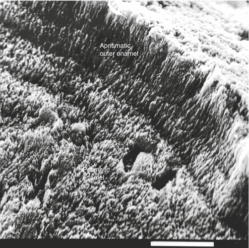
Fig. 2.1
Rat’s molar enamel. (a) Normal aprismatic outer enamel/hydroxyapatite crystals are parallel, forming a continuous palisade-like structure. (b) Beneath the surface, enamel displays a prismatic structure, where prisms (P) (or rods) alternate with interprismatic enamel (IP) (interrod enamel)

Fig. 2.2
Human enamel. The surface outer enamel layer looks laminated. This is not the case for the prismatic enamel located underneath
Remineralization agents induced reduced caries and appropriately promote subsurface deposits. Acid neutralization may be obtained by buffering components of the diet. In addition to an early diagnosis of dental erosion, the detection of isolated factors like sports drinks may be implicated in multifactorial erosions. The significance of the mechanisms involved constitutes prerequisites, which are mandatory to initiate preventive and therapeutic measures.
2.3.1 Four Types of Physical and Chemical Dental Erosions Have Been Reported
Imfeld (1996a, b) has defined different types of tissue losses. According to Ganss (2006), each type of alteration bears its own specificity.
1.
Abrasion is located on the incisal or occlusal surfaces and depends on the abrasiveness of the individual diet. The most abrasive agents are toothpaste, as shown by clinical data and in vitro studies. Both patient factors and material factors influence the prevalence of abrasion.
2.
Attrition results from the action of the antagonistic teeth. It has also been named demastication. It is a mixture of abrasion and attrition.
3.
Abfraction is located mainly in the cemento-enamel region where microfractures occur (also defined as fatigue wear). Wedge-shaped defects are related to abfractions or wedge-shaped non-carious cervical lesions. Analyses that demonstrate theoretical stress concentration at the cervical areas of the teeth have not yet been actually proven. However, there are some strong arguments favoring this possibility. The non-carious stress-induced cervical lesion or abfractions are observed in the buccal surface at the cemento-enamel junction of the teeth, with prevalence ranging in humans from 27 to 85 % (Sarode and Sarode 2013)
4.
Erosion results from the action of acids or chelators acting on plaque-free tooth surfaces. The loss of tooth structure is due to acid dissolution without involvement of bacteria. Regurgitated gastric acids or extrinsic components (soft drinks, acidic fruits) may act as extrinsic agents. Erosion is related to enamel surface softening. Repeated direct removal of a softened enamel layer favors more rapid demineralization and tissue loss (Figs. 2.3b and 2.4b).


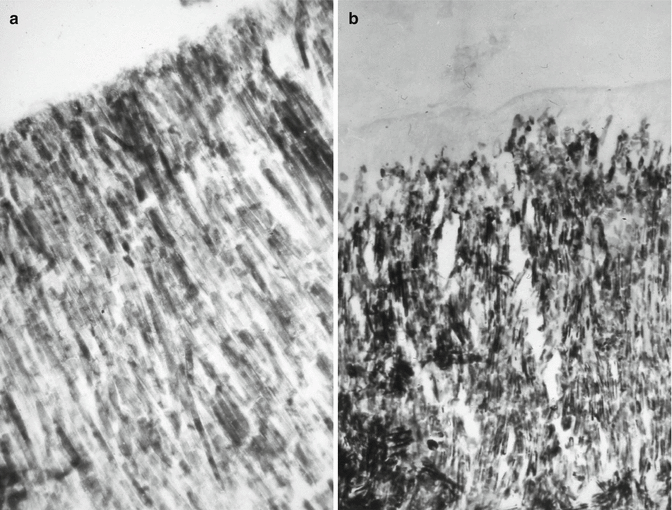
Fig. 2.3
(a) The outer enamel layer displays some interruptions or defects after softening. (b) The surface of corroded enamel displays structural alterations at the tip of crystallites and, at some locations, between crystallites

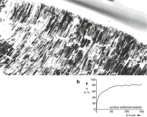
Fig. 2.4
(a) At the surface, crystallite endings look irregular with a scalloped profile. (b) There is a gradual mineral increase after the mineral loss due to the softening agent in the outer enamel surface. The percentage of mineral reaches a “normal” value at 50 μm under the enamel surface
2.3.2 Structure and Chemistry of Dental Erosion
Table 2.1
Table 2.1


Composition percentage in enamel and dentine
Stay updated, free dental videos. Join our Telegram channel

VIDEdental - Online dental courses


