Introduction
The objective of this study was to evaluate the short-term effects of clodronate, a first-generation bisphosphonate, on early alveolar bone remodeling and root resorption related to orthodontic tooth movement.
Methods
The samples consisted of 54 sex-matched Wistar rats (weight, 180-230 g) allocated to the 2.5 mmol/L clodronate, 10 mmol/L clodronate, and control groups (n = 18 for each group). After application of a nickel-titanium closed-coil spring (force, 60 g) between the maxillary central incisor and first molar, 2.5 mmol/L of clodronate, 10 mmol/L of clodronate, or saline solution was injected into the subperiosteum adjacent to the maxillary first molar every third day. All animals received tetracycline, calcein, and alizarin red by intraperitoneal injection at 1, 6, and 14 days, respectively. The amounts of tooth movement were measured at 3, 6, 9, 12, and 15 days. The animals were killed at 4, 7, and 17 days. Histomorphometric analyses of bone mineral appositional rate, labeled surface, percentage of root resorption area, and number of root resorption lacunae of the mesiobuccal root of the maxillary first molar at 4, 7, and 17 days were done. One-way analysis of variance (ANOVA) with the post-hoc test were done for statistical analyses.
Results
Rats in the 10 mmol/L clodronate group had significant decreases of tooth movement (12 and 15 days, P <0.05) and percentages of root resorption area and numbers of root resorption lacunae (7 day, P <0.05), and increases of labeled surface and mineral appositional rates (17 day, P <0.05) over those of the 2.5 mmol/L clodronate and control groups.
Conclusions
Although clodronate might decrease root resorption related to orthodontic tooth movement, patients should be informed about a possible decrease in the amount of tooth movement and a prolonged period of orthodontic treatment.
Recently, orthodontists have been treating aging populations with malocclusion and providing care for esthetic or periodontal problems. These patients have various metabolic bone diseases associated with excessive bone resorption. Identifying the factors related to bone or root resorption after application of orthodontic forces remains a challenge.
Clodronate (dicloromethylene-1,1 bisphosphonate) is a first-generation bisphosphonate and synthetic nonhydrolyzable analog of pyrophosphate. Because it can effectively control the formation and dissolution of calcium phosphate in vitro, as well as mineralization and bone resorption in vivo, it has become a major antiresorptive drug for the prevention and treatment of postmenopausal osteoporosis and various metabolic bone diseases associated with excessive bone resorption. Additionally, it is used to decrease the number and rate of fractures in patients with breast cancer and myeloma-related osteolysis and to relieve metastatic bone pain caused by solid tumors.
In issues related to orthodontic tooth movement, bisphosphonates are known to inhibit root resorption in rats in a dose-dependent manner. Liu et al reported that the number of osteoclasts on the pressure side of the periodontal ligament during orthodontic tooth movement was decreased in animals injected with clodronate. They suggested that the anti-inflammatory properties of clodronate could be helpful in the treatment of periodontitis and that this inhibitory effect was dose-dependent. Other studies demonstrated the inhibitory action of clodronate on bone resorption by apoptosis of osteoclasts through incorporation of clodronate into the cells.
However, to our knowledge, the relationship between the dose of clodronate and early bone remodeling, as well as root resorption during orthodontic tooth movement, has not been proven with histomorphometric studies. Therefore, the purpose of this study was to evaluate the short-term effects of clodronate on early alveolar bone remodeling and root resorption related to orthodontic tooth movement, measured by fluorescent bone markers.
Material and methods
The sample consisted of 54 Wistar rats (approximate weight, 180-230 g; age, 8 weeks) allocated into the 2.5 mmol/L clodronate group, the 10 mmol/L clodronate group, or the control group (n = 18; 9 males and 9 females in each group) to prevent a sex-related bias. This study was conducted under an approved protocol and guidelines established by the Institutional Animal Care and Use Committee of Seoul National University (SNU-090306-4).
After the animals were placed under general anesthesia with zylazine hydrochloride (40 mg/kg, Rompun, Bayer Korea, Seoul, Korea) and zolazepam (5 mg/kg, Virbac, Carros, France), the initial body weights and distances between the tip of the maxillary central incisors and the mesiobuccal cusp tip of the maxillary first molars were measured with a digital caliper (700-126, Mitutoyo, Kawasaki, Japan). Grooves were prepared at the cervical line of the maxillary incisors by using a low-speed hand piece (Vmax, Nakanishi, Tochigi, Japan) to prevent the ligatures from dislodging.
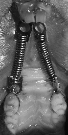
Since several studies demonstrated that 40 to 60 g of force stimulates substantial tooth movement of the molars in rats, a nickel-titanium closed-coil spring (0.10 × 0.30-in; force, 60 g; Ormco, Orange, Calif) was connected with a 0.010-in steel ligature wire between the maxillary first molar and central incisor ( Fig 1 ). After the ligatures were tied, composite resin (Transbond XT Light Cure Adhesive Paste, 3M Unitek, Monrovia, Calif) was cured over the ligature wire to prevent slippage. The mandibular incisors were reduced to prevent breakage of the orthodontic appliance.
Volumetrically equivalent dosages of 2.5 mmol/L or 10 mmol/L of clodronate solution (50 μL; dichloromethylene diphosphonic acid, Sigma, St Louis, Mo) were injected into the subperiosteum area adjacent to the maxillary first molar every third day for the experimental groups with a 31-gauge ultra-fine insulin syringe (BD, Franklin Lakes, NJ) during the experimental period. An equal volume of saline solution was injected into the same area for the control group.
Tetracycline (25 mg/kg, Sigma) on the 1st day, calcein (20 mg/kg, Sigma) on the 6th day, and alizarin red (10 mg/kg, Sigma) on the 14th day were given to all animals by intraperitoneal injection.
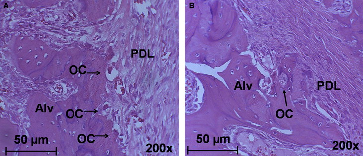
Two animals from each group were killed on the 4th, 7th, and 17th days after spring placement. The specimens of each half of the right and left sides with the adjacent bone tissue were fixed by using 70% ethanol, dehydrated, and embedded in Osteobed resin (Polysciences, Warrington, Pa). The specimens were cut parallel to the mesiodistal long axis of the maxillary first molar and then prepared as 40 to 50 μm thickness specimens by using a cutting and grinding system (Exakt Apparatebau, Nordstedt, Germany). As the observation area, the mesiobuccal root of the maxillary first molar was chosen because it is the largest of the maxillary first molar’s 5 roots and is in approximately the same plane with the applied force.
During the grinding procedure, the specimen was checked with a microscope (SZ 61, Olympus, Tokyo, Japan) to prepare the most suitable slides for histomorphometric analysis. An average of 6 slides for each specimen was obtained; this was sufficient to perform the histomorphometric analysis.
After examination of the fluorescent labeling by using fluorescence microscopy (BX-51, Olympus), the specimens were stained with hematoxylin and eosin.
The amounts of tooth movement were measured on the 3rd, 6th, 9th, 12th, and 15th days after spring placement. Distances between the tip of the maxillary central incisors and the mesiobuccal cusp tip of the maxillary first molars were measured by using digital calipers.
Histologic observation was performed with the Olympus BX-51 microscope ( Figs 2 and 3 ). Bone mineral appositional rate (MAR), labeled surface (LS), percentage of root resorption area (PRRA), and number of root resorption lacunae (NRRL) of the mesiobuccal root of the maxillary first molar were measured and calculated with image-analyzing software (KAPPA, Opto-electronics, Gleichen, Germany).
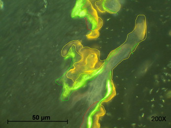
Bone MAR of all the active bone-forming surfaces was determined by measuring the distance between the centers of 2 fluorescent bands (labels) and dividing by the time between labeling (mm/day).
To quantify the overall remodeling activity of the bone at the root surface, LS measurements were made by using spliced images obtained under 200-times magnification. The cumulative lengths of clearly identifiable fluorescent LS were measured between the alveolar bone crest and the apex of the tooth.
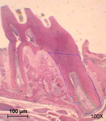
The surface area of the whole root (SAWR, μm 2 ), surface area of root resorption lacunae (SARRL, μm 2 ), and surface area of the radicular pulp (SARP, μm2) were calculated by tracing the pulp area and roots with the image analyzing software ( Fig 4 ). PRRA was obtained by using the formula, SARRL/(SAWR-SARP)] × 100. NRRL measurements were taken from 4 sections at 30-μm intervals and averaged for the mesiobuccal root of the first molar ( Fig 4 ).
One operator (J.C.) did the counting and measuring twice with a 2-week interval. Since there was no significant difference, the first set of counts and measurements was used for analysis.
Statistical analysis
One-way analysis of variance (ANOVA) and a subsequent post-hoc test were performed. P values less than 0.05 were considered statistically significant.
Results
The amounts of tooth movement were compared according to time ( Table I ). Both the 2.5 and 10 mmol/L clodronate groups showed significantly less tooth movement on days 12 and 15 than did the control group ( P <0.05, respectively).
| Day after surgery | Control | 2.5 mmol/L clodronate | 10 mmol/L clodronate | Significance | Multiple comparison test |
|---|---|---|---|---|---|
| 3 | 1.57 ± 0.92 | 1.71 ± 0.81 | 1.61 ± 0.67 | NS | NS |
| 6 | 2.00 ± 0.76 | 2.09 ± 0.91 | 1.96 ± 0.56 | NS | NS |
| 9 | 2.27 ± 0.80 | 1.99 ± 0.56 | 1.90 ± 0.46 | NS | NS |
| 12 | 2.81 ± 0.82 | 2.06 ± 0.50 | 1.93 ± 0.13 | ∗ | (2.5C, 10C) <C |
| 15 | 2.81 ± 0.82 | 1.91 ± 0.50 | 1.79 ± 0.13 | ∗ | (2.5C, 10C) <C |
MAR of the bone was compared near the mesiobuccal root area of the maxillary first molars on days 4, 7, and 17 after spring application ( Table II ). The MAR did not show a significant difference among the 3 groups on days 4 and 7. However, the MAR on day 17 was significantly increased in the 10 mmol/L clodronate group compared with the control and the 2.5 mmol/L clodronate groups ( P <0.05, respectively).
| Day after surgery | Control | 2.5 mmol/L clodronate | 10 mmol/L clodronate | Significance | Multiple comparison test |
|---|---|---|---|---|---|
| 4 | 7.68 ± 4.71 | 6.37 ± 5.16 | 4.90 ± 2.73 | NS | NS |
| 7 | 1.53 ± 0.68 | 4.07 ± 2.28 | 5.05 ± 4.30 | NS | NS |
| 17 | 3.52 ± 1.31 | 3.58 ± 1.12 | 6.62 ± 1.47 | ∗ | (C, 2.5C) <10C |
The LS (μm) of bone was compared near the mesiobuccal root of the maxillary first molars on days 4, 7, and 17 after spring application ( Table III ). There was no significant difference in LS among the 3 groups on days 4 and 7. However, LS in the 10 mmol/L clodronate group was significantly greater compared with that of the control and 2.5 mmol/L clodronate groups on day 17 ( P <0.05, respectively).
| Day after surgery | Control | 2.5 mmol/L clodronate | 10 mmol/L clodronate | Significance | Multiple comparison test |
|---|---|---|---|---|---|
| 4 | 639.86 ± 74.97 | 776.73 ± 267.54 | 845.44 ± 526.88 | NS | NS |
| 7 | 516.67 ± 109.55 | 522.30 ± 89.44 | 513.32 ± 68.28 | NS | NS |
| 17 | 581.87 ± 80.53 | 647.85 ± 179.50 | 978.50 ± 331.75 | ∗ | (C, 2.5C) <10C |
PRRA and NRRL of the mesiobuccal root of the maxillary first molars were compared on days 4, 7, and 17 after spring application ( Tables IV and V ). Although there were no significant differences in PRRA and NRRL among the 3 groups on days 4 and 17, the 10 mmol/L clodronate group showed significant decreases in PRRA and NRRL compared with the control group on day 7 ( P <0.05).


