Introduction
As digital imaging improves and digital cephalometric radiography becomes more prevalent, the need for digital storage space and transmission speed will increase. Compression of the image files is 1 method to overcome transmission overload. However, compression could compromise image quality. The purpose of this study was to determine the range of compression ratios, by using the JPEG2000 standard, within which the identification of landmarks on cephalometric radiographs is not compromised.
Methods
Ten lateral cephalometric digital images were used. Six raters identified 19 landmarks under controlled viewing conditions. The images included the original uncompressed TIFF image and the JPEG2000 format at 3:1, 12:1, 50:1, and 110:1 compression ratios. The images were randomized and displayed with image processing software. The x and y coordinates of each landmark were recorded.
Results
All compression ratios performed equally well compared with the original images with the exception of A-point and nasion at 110:1 and gonion at 3:1 compression ratios. All landmark identifications were precise with the exception of the maxillary incisal apex and edge at the 12:1 and 50:1 compression ratios, respectively.
Conclusions
JPEG2000 is a reliable file format that can be implemented in orthodontic practice.
Digital radiography has been used for a long time in medicine. It was not until the 1980s that the first intraoral sensors were applied in dentistry. The development of cost-effective extraoral digital technology, coupled with increased use of computers in orthodontic practice, has made direct digital cephalometric imaging a valid opportunity. As digital imaging increases, the need for storage space and transmission speed also increases. Digital imaging systems will continue to improve, and, as a result, the storage and transmission requirements for the images they produce will probably increase as well. One method to overcome this transmission overload is to compress the image files. Image compression requires less storage space and decreases the transmission time, since the compressed file is smaller because of the reduced amount of binary data used to represent the image.
There are 2 methods of image compression: lossless and lossy. Lossless compression eliminates nonessential information in the image while conserving essential data so that the digital image can be reconstructed exactly. Lossy compression, on the other hand, although offering considerably higher compression ratios and smaller file sizes, involves irreversible loss of data that could be essential. The most common file format that offers lossy compression is the joint photographic experts group (JPEG) format. The JPEG2000 file format was developed to address some deficiencies in the original JPEG standard. This file format has been adopted by the digital imaging and communication in medicine (DICOM) standard.
Compared with JPEG, JPEG2000 allows higher compression without compromising quality. It also offers progressive image reconstruction, provides more options and greater flexibility than the standard JPEG format, and offers an optional lossless compression mode. Typically, the greater the compression applied to the image to reduce its file size, the lower the image quality. With JPEG2000, there is no artifact with high compression.
Several studies in dentistry have addressed the effects of compression on the diagnosis of caries, periapical lesions, subtraction radiography, and endodontic file length assessment. However, only 2 of these were done with the JPEG2000 standard. The only study that evaluated the effect of compression on lateral cephalometric images used an aluminum test block object to assess the effect of the JPEG compression algorithm on direct digital cephalometric image quality. It was concluded that JPEG compression had no effect on the perceptibility of landmarks in the aluminum test object used in that study.
The purpose of this study was to determine the range of compression ratios, using the newly established JPEG2000 standard, within which landmark identification on cephalometric radiographs is not compromised.
Material and methods
Thirty lateral cephalometric digital images were obtained from the MiPACS Dental Enterprise Solution archiving system (version 3.1, Medicor Imaging, Charlotte, NC). These images had been acquired for routine orthodontic treatment. All radiographic images were acquired with a ProMax (Planmeca Oy, Helsinki, Finland) radiographic machine and exported in uncompressed tagged image file format (TIFF). Those radiographs were selected based on the following inclusion criteria: sharpness of the image, ultimate brightness and contrast, minimal noise, and full visualization of all relevant anatomic structures. From this data set, 10 images were randomly selected.
The inherent resolution of these images was 254 pixels per inch, which is equivalent to 100 pixels per centimeter (1 pixel = 0.1 mm). Image matrix dimension was 2272 × 1818 pixels at 8-bit depth. This resulted in a file size of 4.15 MB. The images were not enhanced in any way.
Photoshop CS2 software (Adobe Systems, San Jose, Calif) was used for compression. Photoshop does not include the JPEG2000 plug-in by default, so it was installed manually. Photoshop uses a quality-factor scale that ranges from 1 (lowest quality) to 100 (highest quality) for JPEG2000 compression. Four JPEG2000 compressed image groups were created by using this software at quality factors of 100, 25, 6, and 1. This produced image file sizes of 1.38 MB, 0.35 MB, 83 KB, and 37.7 KB, respectively. This resulted in compression ratios of 3:1, 12:1, 50:1, and 110:1. The compression ratio expresses the difference between the file size of the original image and the file size of the same image when compressed.
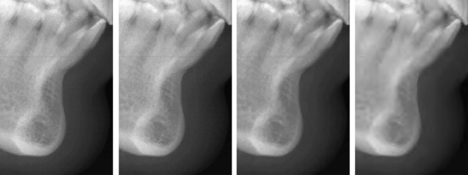
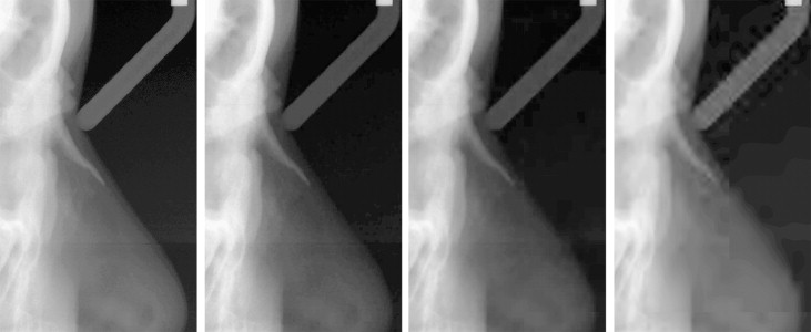
The quality factor of 100 was included in the study because of the reduction in size of the file (one third) and because it was the highest-quality factor that can be achieved by the software. The quality factor of 1 was chosen because it was the lowest quality factor. An example of compressed radiographs is shown in Figure 1 at the mandibular symphysis area. Note in these images how compression at higher levels tends to lose the trabecular bony structure. Another example is shown in Figure 2 at the frontonasal suture and ridge of the nose. Note how soft tissues tend to be affected more than hard tissues by higher compression ratios.


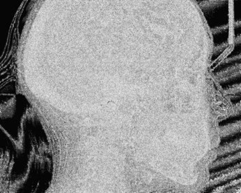
To evaluate the effect of compression at different levels, a cephalometric image at 3:1 compression ratio was qualitatively subtracted from its corresponding uncompressed TIFF image. University of Texas Health Science Center at San Antonio ImageTool software version 3.0 (available on the Internet at http://ddsdx.uthscsa.edu/dig/itdesc.html ) was used for this purpose. The resultant subtracted image indicated that no pixel data had been altered from the original TIFF image. Therefore, the JPEG2000 3:1 compression ratio is lossless. On the other hand, subtraction performed for JPEG2000 images at 12:1, 50:1, and 110:1 compression ratios from the original TIFF showed pixel changes of 59.9%, 69.3%, and 70.2%, respectively. This confirmed that compression at these levels is lossy. According to Figure 3 , most of the pixels that correspond to the patient’s head were changed. It seems that the majority of the 29.8% of unchanged pixels were those that corresponded to air around the patient’s head; these do not change by compression because they are uniformly black and can be compressed efficiently.

Because the ImageTool software used for viewing images does not support the JPEG2000 format, all JPEG2000 images were converted to TIFF. This should not affect the quality of the images; it neither improves nor degrades them. Another subtraction experiment was performed and confirmed this.
The result was 50 images consisting of the original uncompressed images and 4 compression ratios. Randomization of the images was achieved by using a randomizer to blind the viewers.
The images were viewed on an OptiPlex 745 computer (Dell, Round Rock, Tex) with a Core 2 Duo Processor (Intel, Santa Clara, Calif) and the Windows XP professional operating system (Microsoft, Redmond, Wash). The video card was Graphics Media Accelerator 3000 (Intel) using up to 256 MB of system memory. The images were displayed on an UltraSharp 24-in 2007FP TFT flat panel (Dell). Screen resolution was 1920 × 1200 at 60 Hz and 16.7 million colors. The images were presented at 50% zoom because the screen resolution was smaller than the images themselves.
Each image was displayed by using the ImageTool software. For each image, the viewers were asked to identify 19 landmarks commonly identified by orthodontists and previously used in research: N, Or, ANS, PNS, A, B, Pog, Gn, Me, Go, Ar, Po, S, UIA, UIE, LIE, LIA, UM, LM. Definitions of these landmarks are listed in Table I .


