Introduction
In this study, we investigated the nanohardness and elastic modulus of enamel after debonding metal brackets.
Methods
The surfaces of 3 maxillary premolars were subdivided into 3 regions. Two regions were exposed to a conventional etching system (Transbond XT, 3M Unitek, Monrovia, Calif) and a self-etching system (Transbond Plus primer, 3M Unitek); the third region was not etched. Metal brackets were bonded with Transbond XT composite to the 2 etched regions. After storage for 24 hours in distilled water, the brackets and residual adhesive were removed, and the teeth were sectioned transversely. Seven nanoindentations (2 mN load) were placed 1 to 25 μ m from the surface in each region. Mean nanohardness and elastic modulus were compared with analysis of variance (ANOVA) and the Scheffé test.
Results
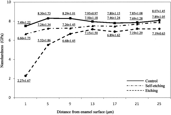
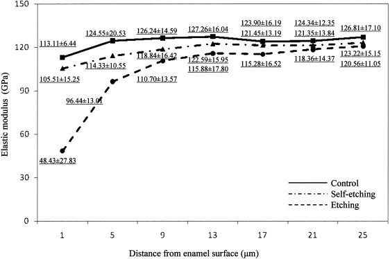
Locations 1 and 5 μ m from the enamel surface had significantly ( P <0.05) lower nanohardness and elastic modulus values for the conventional system compared with the self-etching system and the unetched region. All other locations for the conventional system and all locations for the self-etching system and unetched area had no significant differences. The nanohardness was much higher than the Vickers hardness for enamel.
Conclusions
The minimal effect of the self-etching system on the nanomechanical properties of enamel arises from much lower chemical attack. The much greater effects of the conventional system require further study.
After acid etching was introduced in the mid-1950s, the direct bonding of orthodontic appliances to enamel with epoxy resin was introduced by Newman in the mid-1960s and is now widely accepted by most orthodontists. Bonding practices based on a self-etching primer, which reduces clinical bonding steps by combining etching and priming into 1 step, are now being used in clinical orthodontics. In addition to saving time and reducing procedural errors, their lower etching ability from higher pH compared with phosphoric acid, might minimize the potential for iatrogenic damage to enamel. The mechanical properties of the etched enamel surface should be affected by the degree of demineralization and penetration of the resin tags by bracket bonding. Knowledge of the change in mechanical properties of enamel after debonding brackets is important in understanding iatrogenic damage, such as enamel fracture. Recently, the nanoindentation test has become a common technique for analyzing mechanical properties such as hardness and elastic modulus for small areas and thin regions of materials.
The purpose of this study was to use the nanoindentation test to examine the effects of conventional etching and self-etching on the nanomechanical properties (hardness and elastic modulus) of the enamel surface region. It was hypothesized that self-etching would cause less degradation of these properties than conventional etching. This is the first study that has investigated the nanomechanical properties of enamel after bracket bonding with conventional etching and self-etching systems.
Material and methods
Three noncarious maxillary premolars were used in this study. The teeth had been extracted for orthodontic reasons and with the patients’ informed consent. Selection criteria included no visible decalcification or cracking of the enamel surface under a stereoscopic microscope (SMZ 1500, Nikon, Tokyo, Japan) at magnification of 10 times. The extracted teeth were stored in 0.5% chloramine solution at approximately 4 ° C. The buccal surfaces of the teeth were cleaned by using nonfluoridated pumice. The teeth were subsequently polished by using a rubber cup and thoroughly washed and dried with a moisture-free air source.
The buccal surfaces of the 3 premolars were divided into mesial and distal regions with masking tape (approximately 1 mm width) ( Fig 1 , a ). The mesial region was etched with conventional 35% phosphoric acid gel (Transbond XT Etching Gel, 3M Unitek, Monrovia, Calif) for 15 seconds, washed for 20 seconds, and dried with an oil-free air stream. Transbond XT primer (3M Unitek) was then applied to the etched surface. Transbond Plus self-etching primer (3M Unitek) was next applied to the distal region by rubbing on the enamel surfaces for approximately 3 seconds; afterward, an air jet was lightly applied to the enamel. After the masking tape was removed, metal brackets (Victory Series, 3M Unitek) were bonded with Transbond XT composite resin (3M Unitek) ( Fig 1 , c ) and light-cured for 20 seconds (10 seconds from each proximal side). After storage for 24 hours in distilled water, the metal brackets were debonded, and the residual adhesive was removed by grinding with a low-speed tungsten carbide bur (CA1172, lot 2710291, Shofu, Kyoto, Japan), followed by polishing with nonfluoridated pumice. All teeth were cut with a slow-speed water-cooled diamond saw (Isomet, Buehler, Lake Bluff, Ill) so that they were divided into occlusal and cervical halves ( Fig 1 , f ); 1 sectioned specimen (transverse planes) was then encapsulated in epoxy resin (Epofix, Struers, Copenhagen, Denmark) for the nanoindentation test. All samples were ground with 600-grit sandpaper and polished by using diamond suspensions (3, 1, and 0.25 μ m particle size) to obtain a suitably polished surface.
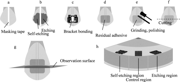
Nanoindentation testing was carried out at 28°C with a nanoindentation system (ENT-1100a, Elionix, Tokyo, Japan) by using a 2-milliNewton (mN) load. The indentations were placed at 49 locations spaced 4 μ m apart (7 × 7 locations; 1-25 μ m depth from the surface) for each of the 3 regions (control, etching, and self-etching regions) on each tooth ( Fig 1 , h ). Nanohardness and elastic modulus were calculated by using the equations in ISO standard 14577.
Statistical analysis
Statistical comparisons were performed with SPSS software for Windows (version 16.0J, SPSS, Chicago, Ill). Mean values of nanohardness and elastic modulus were compared by using 1-way analysis of variance (ANOVA) and the Scheffé multiple range test, with a significance level of 5%.
Results
Mean values and standard deviations for nanohardness and elastic modulus of the polished enamel specimens (transverse planes) are shown in Figures 2 and 3 , respectively. Locations at 1 and 5 μ m from the enamel surface for the conventional etch-and-rinse system had significantly lower nanohardness and elastic modulus values than did other locations. All mean values of nanohardness and elastic modulus obtained for the self-etching system were not significantly different from those for the control specimen. The nanohardness and elastic modulus values were correlated.


