Introduction
The purpose of this study was to evaluate the efficacy of the Schwarz appliance with a new method of superimposing detailed cone-beam computed tomography (CBCT) images.
Methods
The subjects were 28 patients with Angle Class I molar relationships and crowding; they were randomly divided into 2 groups: 14 expanded and 14 nonexpanded patients. Three-dimensional Rugle CBCT software (Medic Engineering, Kyoto, Japan) was used to measure 10 reference points before treatment (T0) and during the retention period of approximately 9 months after 6 to 12 months of expansion (T1). Cephalometric and cast measurements were used to evaluate the treatments in both groups. Also, the mandibular widths of both groups were measured along an axial plane at 2 levels below the cementoenamel junction from a CBCT scan. Differences between the 2 groups at T0 and T1 were analyzed by using the Mann-Whitney U test.
Results
The dental arch (including tooth root apices) had expanded; however, alveolar bone expansion was only up to 2 mm below the cementoenamel junction. There was a statistically significant ( P <0.05) difference between the groups in terms of crown, cementoenamel junction, root, and upper alveolar process. However, no significant ( P >0.05) differences were observed in the interwidths of the mandibular body, zygomatic bones, condylar heads, or mandibular antegonial notches. In the mandibular cast measurements, arch crowding and arch perimeter showed statistically significant changes in the expanded group. The buccal mandibular width and lingual mandibular width values had significant changes as measured from a point 2 mm below the cementoenamel junction.
Conclusions
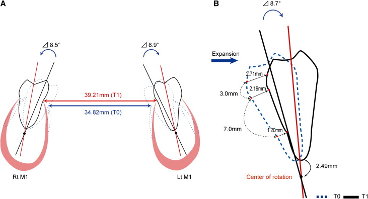
The findings suggest that the Schwarz appliance primarily affected the dentoalveolar complex, but it had little effect on either the mandibular body or any associated structures. In addition, the molar center of rotation was observed to be below the root apex.
Editor’s comment
The treatment effect of orthodontic appliances is frequently debated, mainly because of the lack of clinical evaluation studies. This article provides evidence regarding the dental and skeletal changes induced by the Schwarz appliance in the mandibular arch. The authors used CBCT images to measure tooth and alveolar bone expansion at different levels: the crown of the tooth, 3 mm above the cementoenamel junction (CEJ), the CEJ, 2 mm below the CEJ, and 13 mm below the CEJ. They found that the dental arch expanded by controlled tipping of the posterior teeth, with the center of rotation approximately 2 mm below the root apex of the molars. No skeletal expansion was found. They did not assess the stability of this treatment, only the immediate effects from the appliance.
The study is described as preliminary because the sample size was small; however, the results clearly lead to the stated conclusions. Future studies could expand on the sample size, but the methodology might need to be modified for ethical reasons. Concerns about radiation hazards from CBCT imaging are making it increasingly difficult to obtain approval, especially in studies with children and imaging at several times. The American Association of Orthodontists and the American Association of Maxillofacial Radiology are working on a joint position paper on imaging ( http://www.aaomembers.org/MyPractice/Technology/AAO-Imaging-Initiative.cfm ), and the SEDENTEXCT project ( www.sedentexct.eu/home ) of the European Atomic Energy Community (Euratom) is currently establishing guidelines specifically for CBCT in dentistry.
Demetrios J. Halazonetis
Kifissia, Greece


