and Jasdeep Kaur1
(1)
Earth and Life Sciences Vrije Universiteit Amsterdam and ILEWG, Amsterdam, The Netherlands
13.1 Introduction
13.5.1 Lens Aperture
13.5.2 Depth of Field
13.5.3 Shutter Speed
13.5.4 Film Speed
13.5.5 Focus
13.5.6 Lenses (Angle)
13.8.1 Visible Light Photography
13.8.2 Alternate Light Photography
13.8.4 Infrared Light Photography
13.9 Conclusions
Abstract
Forensic photography regularly represents the paramount method to collect and preserve evidence in forensic cases including forensic odontology. Modern technology enables the investigator to obtain photographs using wavelengths of the light spectrum that are normally obscured from vision. The use of ALI, UV, normal, and IR light source photography for the documentation of the examination, latent fingerprinting processing, detection of semen and bloodstains, trace wound pattern detection, teeth restorations, and bite mark documentation has opened a new frontier for forensic science. This chapter discusses the types of photographic evidence and the specific techniques utilized in full spectrum forensic digital photography.
13.1 Introduction
Physical evidence documentation through forensic photography remains one the most vital features of crime scene investigation. The subsequent analysis of photographs will often yield clues investigators can use to recreate the stages of an event, or may provide the proof necessary to gain a conviction at trial. Conventional photography records images in the visible light spectrum and typically will record on film or in a digital file that the human eye can see. Forensic odontogist photographers are often confronted with evidence where traditional photographic techniques are unsuccessful at documenting the evidence necessary to settle the facts of a legal case. For years forensic photographers have had a variety of specialized techniques available for documenting evidence under challenging situations. Infrared, ultraviolet (UV), and alternate light imaging (ALI) photographs can be used in a variety of these situations to gain results that could not be obtained by photographing in the visible light spectrum. A forensic light source is a crime scene investigator’s and lab technician’s tool for enhancing observation, photography, and collection of evidence, including latent fingerprints, body fluids, hair and fibers, bruises, bite marks, wound patterns, shoe and foot imprints, gunshot residues, drug traces, questioned documents, bone fragment detection, and so on (Wright and Dailey 2001; Wright and Golden 1997). Thus, it is essential that the forensic odontologist accountable for the evidence collection understands the capabilities of full spectrum digital photography. Different light forensic photography is very important in cases involving dental identification, human abuse, and bite marks. The purpose of this chapter is to review the application of forensic light sources in dental legal cases and other cases.
13.2 Basic Concept of Light and Spectrum of Light
The concept of color and photoluminescence emission stands for the observation of physical phenomena due to the interaction of light with material. Light consists of electromagnetic energy, including microwaves, radio waves, etc. The visible waves, which make up white light, form only a part of the electromagnetic spectrum. The capability of our eyes to see visible light is called color vision and is similar to a filter that transmits only the radiation between approximately 400–700 nm (Fig. 13.1). Radiation at 420 nm is observed as blue, at 450 nm as alternate light or yellow, at 550 nm as green, and at 650 as red. The mixture of electromagnetic rays between 400 and 700 nm is seen as white light.


Fig. 13.1
Spectrum of light
13.3 Electromagnetic Spectrum and Skin
Photographic techniques utilizing these four types (visible light, ALI light, UV light, and IR light) of the light spectrum give very different looks into pattern injuries on skin. Because the human eye is unable to see beyond the visible light spectrum, these special types of photographic techniques are applied to produce images in the nonvisible zones of electromagnetic radiation such that they can be seen with the human eye. When light hits human skin, different types of processes take place, including reflection, absorption, fluorescence, and scattering of the light. Reflection occurs as the shorter wavelengths of light strike the surface of the skin. Depending on different parameters of radiation, more than 60% of short wavelengths do not penetrate the surface of the skin and are reflected back (Anderson and Parrish 1982; Rai and Kaur 2010), while the longer wavelengths of light (700–900 nm) can penetrate the skin up to 2–3 mm (Anderson and Parrish 1982; Rai and Kaur 2010). When light rays strike the skin, a molecular-level excitation within the skin occurs that increases the resting state energy of the molecules within the skin, known as fluorescence. This phenomenon remains for about 100 ns. A special technique known as alternative light imaging is needed for the human eye to see and the camera to record this type of fluorescence (Wright 1998). This technique is named for Prof. G. G. Stokes, who reported in 1853 that the remitted wavelength of a predominant color of light is of a different frequency than the illuminating source. A part of the energy of the light at a particular frequency is absorbed by the subject matter it strikes. Once that energy is absorbed in the form of electrons, it creates a molecular excitation that looks for a return to its rest position. The return of the electrons to their resting position releases that energy as fluorescence (Stokes 1853).
13.4 Basic Concept of Photography, Inflammation, and Repair
It is most important for forensic photographers to understand the physiological changes that take place during a skin injury. These changes from a normal state to an injured state to a healing state permit us to differentiate among the contusions illuminated by light sources of various wavelengths. A skin injury leads to inflammation and repair (Wright and Golden 1997, 2010). Inflammation leads to vascular changes at the site of the injury, as the body starts to transport blood in response to tissue damage and to mediate the framework for healing. The damaged tissues further lead healing through the process of repair or scarring. The tissues heal by repair via cell replication, while if damage is too large for repair, the tissue will heal by scarring. Inflammation is the strongest soon after the injury occurs, with repair becoming the prevalent event as the tissue begins to heal. The chemical composition of the injured skin is very different from the surrounding normal, healthy skin. Damaged tissues respond a different way to electromagnetic radiation than the adjacent normal, healthy skin because of differences in photoactive agents, including hemosiderin, melanin, hemoglobin, beta-carotene, etc. (Wright and Golden 1997).
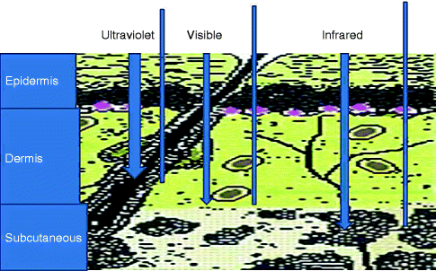
When the pattern injury in the skin shows signs of injury on the epidermis, it is best to use the UV range to photographically illustrate the surface damage because it allows more absorption and has a shallower depth of penetration (Wright and Golden 1997, 2010). UV photography requires a special armamentarium for taking UV photographs. If a skin injury shows signs of an injury on the dermis, it would require using longer wavelengths of light that could go through the skin to the level of the bruising, namely, longer wavelengths of light in the IR range. It is very important to distinguish between the healthy adjacent skin and the injured skin. It would require using a special type of photography known as fluorescent photography techniques (ALI) (Wright and Golden 1997). All photography requires a totally dark room. The forensic photographer can employ any or all of the photographic techniques to take and preserve the evidence.
Note: Visible light photography creates images of the injuries as seen by the unaided eye. UV photography shows details of the damaged epidermis surface of the skin, while IR photography exhibits the tissue injury at the dermis and below. ALI photography will record the difference between the uninjured skin adjacent to the injured skin using fluorescence (Fig. 13.2). It is important to understand that all techniques will create useful images at all times (Rai and Kaur 2010).


Fig. 13.2
Penetration of different light wavelengths in skin
13.5 Different Parameters of Photography (Redsicker DR 1994)
The light can be controlled three ways:
1.
Lens aperture (f-stops), e.g., f/2 (maximum light), f/2.8, f/4, f/5.6, f/8, f/11, f/16 (minimum light)
2.
Shutter speed, e.g., 1/(maximum light) … 1, 2, 4, 8, 15, 30, 60, 125, 250 (minimum light)…
3.
Speed of film (ISO/ASA), e.g., 64 (less light), 100, 200, 400, 800, 1,600 (more light)
Digital cameras are becoming entirely automatic. This does not make them foolproof; it just means they are more complicated to use appropriately. Be familiar with your camera—read your manual—have your manual with you—refer to it.
13.5.1 Lens Aperture
The lens aperture ring limits the amount of light allowed through the lens. This limiting is selected with f-stops—numerical representations of the aperture, where the number increases as the aperture (opening) decreases:
Notes
(a)
Large opening (small no.), small opening (large no.).
(b)
Light requires less light/more light?
(c)
Depth of Field: narrow/wide?
(d)
Place your subject at a far enough distance so that your lens indicates that the maximum depth of field extends to infinity. Therefore, your “sharpness” extends from your minimum range of depth of field (designated on your lens) to infinity; thus, you achieve maximum depth of field.
Stay updated, free dental videos. Join our Telegram channel

VIDEdental - Online dental courses


