Fig. 5.1
Effect of the continuous exposure of a human third molar to 10 % citric acid. The amorphous, centripetal tissue loss is obvious: (a) unaffected tooth, (b) tissue loss after 12 h immersion time. In part from Ganss [1]
The diagnostic criteria for dental erosion were derived from early observations in small groups of patients with known acid exposures [3, 4], and are meanwhile established in clinical diagnosis [5] and erosion index systems [6]. This type of tooth wear is acid-mediated with no bacteria involved. In initial stages of lesion development, the tooth surface may appear dull and the normal surface structures of enamel flatten. In later stages, a distinct defect develops. On occlusal surfaces cusps are cupped, the morphology flattens and restorations may stand proud of the surface (Fig. 5.2). Normally, there are no occlusal contacts at the lesion area. Finally, the crown height may be severely reduced. On smooth surfaces, erosive lesions occur coronal from the enamel-cementum junction often leaving an intact cervical rim (Fig. 5.3). The lesions are shallow and can affect the entire tooth surface.
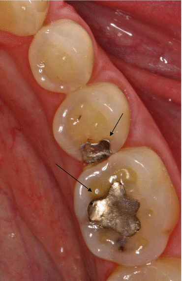
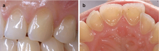
Fig. 5.2
Occlusal erosion. Typical signs are flattening of the occlusal morphology, cupping of the cusps and filling proud of the surface (arrows)
Fig. 5.3
Erosion on smooth surfaces. (a) Shallow lesions coronal from the enamel-cementum junctions at the buccal surfaces. Note the intact enamel rim at the gingival margin. (b) Palatal erosion: the palatal morphology has disappeared, note also the cervical intact enamel rim
The differential diagnosis of erosive wear is tissue loss predominantly caused by physical forces namely attrition, abrasion and abfraction/wedge-shaped defects (non-carious cervical lesions).
Attrition is the result of two-body wear from tooth-to-tooth contacts. Respective lesions (Fig. 5.4a) can only occur in occluding areas of the dentition, normally on occlusal surfaces. Its typical shape is flat and sharply bordered and its surface appears highly polished. Typically, lesions occur on both opposing tooth surfaces (Fig. 5.4b).
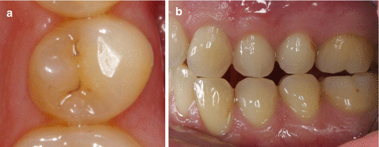
Fig. 5.4
Attrition. (a) Typical flat, sharp-bordered tissue loss on the buccal cusp of a second upper premolar. (b) The same subject as in (a) – note the attritional defects on opposing teeth
Abrasion occurs as result of three-body-wear, e.g. from toothbrushing with toothpaste or the interaction of opposing teeth with a food bolus (demastication). This form of tooth wear occurs mainly in dentin which is much less wear-resistant than enamel [7]. On smooth surfaces, abrasion thus is mainly located apical to the enamel-cementum junction and occurs on exposed root surfaces (Fig. 5.5). On occlusal surfaces, exposed dentin is hollowed out resulting in cupping of cusps and grooving of incisal edges (Fig. 5.6a). These lesions look very similar to occlusal erosive lesions (Fig. 5.6b). Abrasive lesions are smooth-bordered, and can be of variable shape depending on the causing impact.
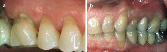
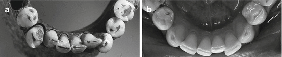
Fig. 5.5
Abrasion on smooth surfaces. Lesions are located apical to the enamel-cementum junction, combined with gingival recession. (a) Lesions at teeth 16–14, 65-year-old woman. (b) Lesions at 23, 24, 26, 33 and 32, 30-year-old woman. Note that lesions at the first upper molars look very similar and would be regarded as a more severe stage at younger than at older age
Fig. 5.6
Abrasive lesions on occlusal and incisal surfaces (demastication) in a medieval remain (a) resulting from an abrasive diet and in a subject with chronic vomiting (b). Note the strikingly similar shape of tissue loss (in part from Ganss [1])
Abfraction/wedge-shaped defects (also called non-carious cervical lesions) are located at the enamel-cementum border. Its coronal part cuts more or less rectangular into the enamel and normally is sharply demarcated, whereas its apical part runs flatly towards the root surface (Fig. 5.7). The aetiology of this type of lesions is still not clarified. It was suggested to be the result of either improper toothbrushing or eccentric functional forces causing flexural stress at the cervical area of the tooth disaggregating the enamel crystals, or an interaction of both [8]. Non-carious cervical lesions and its management is discussed in detail in Chap. 14 of this book.
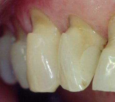
Fig. 5.7
Wedge-shaped defect (abfraction): the lesion is located at the enamel-cementum junction. It cuts more or less rectangular into the enamel at its coronal part and flattens towards the root surface at its apical part
The differential diagnosis on smooth and occlusal surfaces is illustrated in Figs. 5.8 and 5.9. The most challenging situation is to distinguish between abrasive and erosive lesions on occlusal/incisal surfaces because both types of tissue loss may look very similar (Fig. 5.6a, b). This was shown in a study investigating tooth wear in subjects with substantially different nutrition [9]. Lesions on occlusal surfaces in medieval remains with an abrasive diet and in contemporary subjects with an acidic diet were more severe in the former, but were similar in shape; incisal grooving was a common condition in all subjects and was not related to any form of diet. Strikingly, smooth surfaces were never affected in subjects with abrasive diet, but frequently exhibited shallow lesions coronal to the enamel-cementum junction in subjects with acidic diet. The conclusion from this study is that such shallow smooth surface lesions may be a reliable criterion for distinguishing between abrasive and erosive tooth wear.
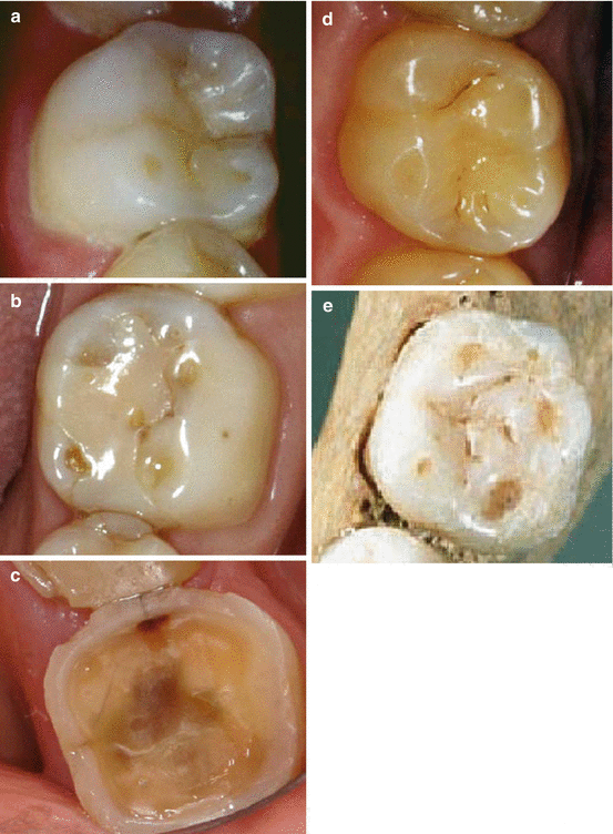
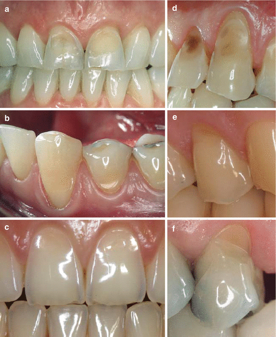
Fig. 5.8
Differential diagnosis of occlusal tooth wear. (a–c) Initial, moderate and severe erosive lesions. (d) Attrition at the mesio-palatal cusp with the typical flat and sharp-bordered shape. (e) Abrasion (demastication) in a medieval remain, note the strikingly similar shape of tissue loss compared to (b)
Fig. 5.9
Differential diagnosis of tooth wear on smooth surfaces. (a–c) Erosive lesions of buccal surfaces mainly located coronal from the enamel-cementum junction with an intact cervical enamel rim. (d) Abrasion typically smooth-bordered and located mainly on the root surface. (e, f) Wedge-shaped defects (abfraction) typically located at the enamel-cementum junction
In the individual subject, the clinical differential diagnosis of tooth wear can be substantiated by a thorough anamnesis and by nutrition diaries [5]. The anamnesis should cover the variety of aetiological factors of tooth wear concerning diet, oral hygiene, hobbies, sports and occupation and should address not only current, but also earlier conditions (See Chaps. 3 and 4). Many patients are not aware about the erosive potential of food and drinks, thus nutrition diaries may help identifying acidic food and drinks as well as other nutritional factors contributing to tooth wear. Dietary management in dental erosion is discussed in detail in Chap. 10 of this book.
5.3 Defining the Stage of Dental Erosion and Assessing Progression
At any stage, dental erosion ceases from progression as soon as the causal factor is sufficiently limited. However, when a clinically visible defect has developed, there is no option for restitutio ad integrum and lost dental hard tissue cannot regenerate. Different stages of erosive tissue loss require adapted preventive and therapeutic strategies [10]. Diagnosing early stages is important to initiate appropriate preventive measures to avoid further tissue loss. More advanced tissue loss may require a more complex intervention and in severe cases the lost tissue may need to be restored.
Stay updated, free dental videos. Join our Telegram channel
VIDEdental - Online dental courses


