Introduction
The aims of this study were to evaluate the in-vivo reaction of newly erupted enamel to demineralization around orthodontic brackets and to compare it with that of mature enamel.
Methods
Thirteen orthodontic patients scheduled to have 4 first premolars extracted for orthodontic reasons were divided into 2 groups. Group 1 included 7 younger patients with newly erupted teeth (4 boys, 3 girls; mean age, 11.21 ± 1.12 years; range, 11-13 years). Group 2 contained 6 adults with mature teeth (5 men, 1 woman; mean age, 34.64 ± 4.01 years; range, 25-41 years). Brackets were placed, and, 30 days later, the teeth were extracted. These teeth were longitudinally sectioned, and demineralization was assessed by cross-sectional microhardness. Determinations were made at the bracket-edge composite limits and at occlusal and cervical points 100 μm away. Evaluations under the brackets and at the lingual surfaces were made as controls. In all these positions, 6 indentations were made at depths from 10 to 90 μm from the enamel surface. Analysis of variance (ANOVA) and Tukey tests were used for statistical evaluation at the P <0.05 level.
Results
ANOVA showed statistically significant differences for tooth type, position, depth, and their interactions ( P <0.05), except the tooth type and position interaction. The multiple comparison test showed less demineralization in the enamel around orthodontic brackets bonded to mature teeth campared with newly erupted teeth ( P <0.05).
Conclusions
During the 30-day study period, the tooth enamel in the adult orthodontic patients was more resistant to demineralization than that of the younger patients.
One of the most difficult problems in orthodontic treatment with fixed appliances is the control of enamel demineralization around brackets. The bands and brackets and the various orthodontic elements (elastics, power chains, sleeves, springs) make the patient’s oral hygiene more difficult and the accumulation of plaque easier. Studies have documented significant increases in oral bacteria during orthodontic treatment. Demineralization takes place when specific bacteria are retained for a long time on the enamel surface. Patients with fixed orthodontic appliances have an elevated risk of caries, and enamel lesions can occur within a month, irrespective of mechanical plaque control and whether fluoridated dentifrices are used. Previous studies have shown that the rate of demineralization in orthodontic patients is higher than in those without orthodontic treatment, and teenagers have a higher risk of demineralization than do adults.
Odontogenesis is a complex process: a series of events from bud formation until the completion of calcification and the maturation of the tooth. Upon eruption, the outermost layer of enamel is immature and not completely calcified, and it then starts to calcify from the effects of salivary minerals. Because of wear and replacement of organic material by minerals during the maturation process, the enamel surface of old teeth might have a different composition than that of newly erupted teeth.
In newly erupted teeth, high sodium and magnesium levels are thought to contribute to the relatively high solubility of enamel. Mineral content in the enamel surface is transformed mainly to calcium phosphate, with little sodium and magnesium remaining during posteruptive maturation. These changes are thought to occur mostly during the first few years after eruption. It was shown in the literature that decreases in enamel pore size and increases in the calcification of enamel matrix occur over time. Also, during aging, hydroxyapatite crystals increase in size because of the incorporation of ions from the surrounding saliva. This is attributed to the mineralization of calcium and phosphate ions from saliva and bacterial acid products.
Imanishi and Nishino showed altered surface and subsurface enamel in newly erupted premolars when compared with premolars from 18-year-olds. Palamara et al used scanning electron microscopy to study the superficial and deep layers of enamel from unerupted and erupted teeth. They also found marked differences in the unerupted surface enamel structure compared with erupted enamel. All these changes during the posteruptive maturation might influence the etching, bonding, and demineralization properties of newly erupted and mature enamel.
Previous studies on posteruptive maturation and its effects on caries development showed decreases in the incidence of caries as the subjects age, confirming the continuous mineralization and maturation of the enamel. The structural characteristics of enamel and their role in the bonding mechanism have been studied previously, but no in-vivo studies have investigated the demineralization properties of newly erupted and mature teeth around orthodontic brackets.
Therefore, the aims of this study were to evaluate the in-vivo reaction of newly erupted enamel to demineralization around orthodontic brackets and to compare it with mature enamel quantitatively. In this study, the null hypothesis assumed that newly erupted enamel showed significantly higher demineralization around orthodontic brackets than do the mature teeth evaluated in the mouth.
Material and methods
This study was approved by the Ethical Committee on Research of the Gulhane Military Medical Academy, Ankara, Turkey. Thirteen orthodontic patients, scheduled to have 4 first premolars extracted for orthodontic reasons, were invited to participate in the study, and consent forms were signed. A power analysis established by G ∗ Power software (version 3.0.10, Franz Faul Universität, Kiel, Germany). Based on a 1:1 ratio between groups, a sample size of 13 patients would give more than 80% power to detect significant differences with 0.40 effect size and at the α = 0.05 significance level. For group standardization, before starting the study, all patients’ teeth were evaluated clinically and radiographically to determine the baseline caries risk. They were divided into 2 groups according to age.
Group 1 (newly erupted teeth) included 4 boys and 3 girls (mean age, 11.21 ± 1.12 years; range: 11-13 years); group 2 (mature teeth) included 5 men and 1 soman (mean age, 34.64 ± 4.01 years; range, 25-41 years).
The patients’ salivary flow rates and buffer capacities were recorded. The criteria for including the patients were no active caries lesions, developmental defects, or fluorosis; and normal salivary flow rate (>1.0 mL/min) and buffer capacity (final pH, 6.5-7.2). All patients received a full-mouth cleaning to remove plaque in preparation for bonding. For evaluating the baseline demineralization values of all selected teeth, a portable battery-powered laser fluorescence device, DIAGNOdent Pen (KaVo Dental, Biberach/Riβ, Germany) was used. The 2 groups’ scores were less than 13, indicating no demineralization; they were equivalent for caries risk.
Orthodontic brackets were bonded with Transbond XT (3M Unitek, Monrovia, Calif), a resin-based composite. A 37% phosphoric acid gel (3M Dental Products, St Paul, Minn) was used for 15 seconds. The teeth were rinsed with water for 30 seconds and dried with an oil-free source for 20 seconds. Transbond XT primer (3M Unitek) was applied to the etched surface in a thin film and not cured. Adhesive paste was applied to the bracket base (Dyna-Lok series, 100-gauge mesh, 3M Unitek), and the bracket was positioned on the tooth and pressed firmly into place. The excess adhesive was removed around the bracket with a scaler, and the adhesive was light cured from the mesial and distal aspects for 10 seconds each (total time, 20 seconds). A light-emitting diode unit (Elipar Freelight 2, 3M ESPE, St Paul, Minn) was used for curing the specimens.
Twenty-eight brackets were bonded in group 1 (14 maxillary and 14 mandibular first premolars), and 24 brackets were bonded in group 2 (12 maxillary and 12 mandibular first premolars). After 30 days, the teeth were extracted and stored in a refrigerator in flasks containing gauze dampened with 2% formaldehyde (pH 7.0) until the analysis. Demineralization in enamel around the brackets was evaluated with the cross-sectional microhardness method according to the literature. During the experimental period and 3 weeks previously, all subjects brushed their teeth with a nonfluoridated dentifrice. They received no instructions regarding oral hygiene, kept their usual habits, and were instructed not to use any antibacterial substance.
One operator (S.O.), blinded to the group allocations, evaluated demineralization. The roots were removed 2 mm apical to the cementoenamel junction, and the crowns were hemisectioned vertically into mesial and distal halves with a 15 high cut (large) wafering blade on an Isomet low-speed saw (Buehler, Lake Bluff, Ill) directly through the slot of the bracket, leaving gingival and incisal portions. The teeth were embedded in self-curing EpoKwick epoxy resin (Buehler), leaving the cut face exposed. The half-crown sections were polished with 3 grades of abrasive paper discs (320, 600, and 1200 grit); final polishing was done with a 1-μm diamond spray and a polishing cloth disc (Buehler). A microhardness tester (HMV-700, Shimadzu, Kyoto, Japan) under a 2N load for 15 seconds was used for the microhardness analysis.
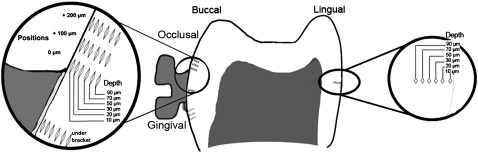
Thirty-six indentations were made on each half crown at 6 positions ( Fig ). On the buccal surface, the first indentations were made under the bracket. In the occlusal and cervical regions, the indentations were made at the edge (0 μm) of the bracket and 100 μm away. Indentations were also made in the middle third of the lingual surface of each half crown as another control. In all these positions, 6 indentations were made at depths of 10, 20, 30, 50, 70, and 90 μm from the external surface of the enamel. These values in the 2 half crowns were averaged.
Statistical analysis
Data analyses were performed with the Statistical Package for Social Sciences, (version 13.0, SPSS, Chicago, Ill) and Excel 2007 (Microsoft, Redmond, Wash). The Shapiro-Wilks normality test and the Levene variance homogeneity test were applied to the demineralization data. The data showed normal distribution, and there was homogeneity of variances between the groups.
Analysis of variance (ANOVA) was used to evaluate the effect of tooth type (newly erupted and mature), depths from the enamel surface (10, 20, 30, 50, 70, and 90 μm), positions (under the bracket, on the buccal surface in the occlusal and cervical regions at 0 and 100 μm from the brackets, and on the lingual surface), and their interactions. For multiple comparisons, the Tukey post-hoc test was used. Significance was predetermined at P <0.05.
For evaluating intraobserver and interobserver agreement, the demineralization measurements were made by 2 investigators (S.O. and T.U.) using the same instrument at 2 separate times, and the Cohen kappa scores were determined.
Results
The kappa scores for the assessment of intraexaminer and interexaminer agreement were higher than 0.80; this implies substantial agreement between the observers.
ANOVA showed statistically significant difference for the factors of tooth type, position, and depth ( P <0.05). The interactions (tooth type/depth, and position/depth) and (tooth type/position/depth) were also statistically significant ( P <0.05) ( Table I ). ANOVA to evaluate the interactions of demineralization for tooth type at various positions under, occlusal, and cervical to the brackets on the labial and lingual (control) surfaces indicated no statistically significant interaction between tooth type and position ( P >0.05) ( Table I ). All descriptive statistics of demineralization at the various depths and positions for newly erupted and mature teeth are shown in Table II . The lowest demineralization values were determined at the occlusal (264.920 VHN ± 16.533) and cervical (253.562 VHN ± 15.129) margins (at 0-μm position) at 10-μm depths for newly erupted premolars.
| Source | Sum of squares | df | Mean square | F | Significance |
|---|---|---|---|---|---|
| Tooth type (newly erupted/mature) | 10110.297 | 1 | 10110.297 | 76.722 | 0.000 ∗ |
| Position | 119532.316 | 6 | 29883.079 | 226.767 | 0.000 ∗ |
| Depth | 1716418.300 | 5 | 343283.660 | 2605.001 | 0.000 ∗ |
| Tooth type/position | 191.710 | 6 | 47.928 | 0.364 | 0.835 |
| Tooth type/depth | 3001.374 | 5 | 600.275 | 4.555 | 0.000 ∗ |
| Position/depth | 103733.489 | 30 | 5186.674 | 39.359 | 0.000 ∗ |
| Tooth type/position/depth | 2653.311 | 30 | 132.666 | 5.007 | 0.000 ∗ |
| Cervical 100 μm | Cervical 0 μm | Under Bracket | Occlusal 0 μm | Occlusal 100 μm | ||||||||
|---|---|---|---|---|---|---|---|---|---|---|---|---|
| Tooth type | Depth | n | Mean (VHN) | SD | Mean (VHN) | SD | Mean (VHN) | SD | Mean (VHN) | SD | Mean (VHN) | SD |
| Newly erupted | 10 μm | 28 | 289.948 | 14.560 | 253.562 | 15.129 | 311.560 | 9.710 | 264.920 | 16.533 | 276.770 | 9.654 |
| 20 μm | 28 | 299.947 | 14.707 | 270.477 | 14.351 | 320.437 | 10.412 | 283.477 | 13.860 | 295.947 | 9.149 | |
| 30 μm | 28 | 320.905 | 13.676 | 292.382 | 12.033 | 332.872 | 8.532 | 304.382 | 12.921 | 307.977 | 9.260 | |
| 50 μm | 28 | 346.367 | 10.190 | 344.737 | 10.190 | 348.367 | 8.330 | 344.737 | 10.190 | 349.367 | 8.908 | |
| 70 μm | 28 | 364.178 | 10.534 | 359.740 | 9.765 | 365.687 | 9.299 | 359.740 | 10.340 | 369.000 | 8.473 | |
| 90 μm | 28 | 369.082 | 9.931 | 369.632 | 8.514 | 370.342 | 8.665 | 369.632 | 8.891 | 371.796 | 7.371 | |
| Mature | 10 μm | 24 | 300.333 | 14.691 | 265.910 | 17.167 | 318.333 | 8.460 | 272.910 | 17.822 | 286.343 | 17.213 |
| 20 μm | 24 | 314.026 | 13.488 | 276.326 | 15.659 | 324.286 | 8.684 | 291.226 | 13.344 | 297.496 | 14.606 | |
| 30 μm | 24 | 322.321 | 12.679 | 298.031 | 13.522 | 339.711 | 8.092 | 312.821 | 11.708 | 317.561 | 12.374 | |
| 50 μm | 24 | 348.606 | 10.021 | 350.226 | 13.008 | 352.916 | 7.379 | 348.156 | 11.363 | 357.916 | 12.773 | |
| 70 μm | 24 | 368.806 | 9.170 | 363.026 | 10.021 | 363.186 | 8.399 | 361.461 | 10.208 | 367.656 | 9.170 | |
| 90 μm | 24 | 369.600 | 7.932 | 370.190 | 9.762 | 373.340 | 9.762 | 374.040 | 9.765 | 374.410 | 8.902 | |
Stay updated, free dental videos. Join our Telegram channel
VIDEdental - Online dental courses


