Fig. 2.1
(a) Midline diastema: space between maxillary central incisors, (b) polydiastema: generalized spacing between teeth
Generally these spaces create an unpleasant appearance for individuals. Sometimes they may lead to phonetic problems, particularly in cases with wide spaces [2].
Diastema necessitates treatment because of esthetic, psychological, and functional concerns. However, the maxillary midline diastema is a normal growth feature of children in the primary and mixed dentition period and, in most of the cases, it decreases or even completely closes by the medial eruption of the maxillary lateral incisors and canines in childhood [2–5]. However, for some individuals, the spaces remain after the transition of dentition [2, 6]. In contrast to the maxillary diastema, the mandibular one is rarely seen in children and is more dramatic than the maxillary diastema. To date, it has been reported that not only a factor is responsible in the formation of diastema but it has multifactorial etiology including the possible genetic predisposition [2, 3, 5, 7–9]. Due to the multifactorial etiology of the diastema, it is important to understand the causes of the condition to select the most appropriate treatment.
Key Note
Due to multifactorial origin of the diastema, clinician should understand the etiological factor before initiating any treatment modality.
2.1 Causes of Congenital or Acquired Diastema
2.1.1 Physiological Development of the Dentition
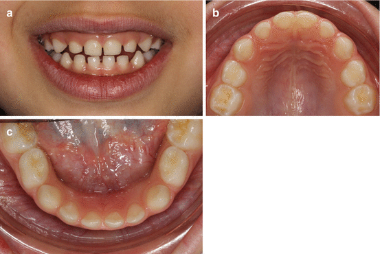
As mentioned above, spacing in anterior teeth is a normal feature of the primary and mixed dentition. The spacing of the primary dentition is the sign of the available spaces allowing for accommodation and proper alignment of the permanent teeth which are larger in size than their preceding [10, 11]. The average primary interdental spacing is 4 mm in the maxilla and 3 mm in the mandible [10] (Fig. 2.2a–c). This physiological diastema normally decreases or closes by the eruption of the lateral incisors and/or canines in most of the cases [2–5, 12]. If there is no blocking pathologic or physiologic condition, spontaneous closure can be expected [5].


Fig. 2.2
(a–c) Physiologic spacing in a 5-year-old girl required for the proper alignment of the permanent incisors
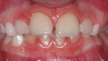
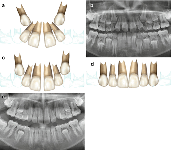
Maxillary midline diastema may persist until the end of the mixed dentition [13] (Fig. 2.3). In this stage of development, also referred as “ugly duckling stage,” developing canines mesially push the roots of the central and lateral incisors causing distal movement of the crowns, leading to maxillary midline diastema (Fig. 2.4a–e). If the midline spacing is 2 mm or less, it will be spontaneously corrected by the eruption of the permanent canines resulting in mesial movement of the incisor’s crowns, whereas a greater diastema is unlikely to close without intervention [14].


Fig. 2.3
Maxillary midline diastema in mixed dentition stage
2.1.2 Heredity and Ethnicity
A familial incidence of diastema has been shown, indicating a hereditary and genetic disposition [7, 9]. Its hereditary background is probably due to the genetic control of the tooth size and agenesis which are the most common etiological factors of dental spacing and seems to be supported by studies revealing the genetic basis of hypodontia and microdontia [15, 16]. Analysis of a pedigree data suggested an autosomal dominant mode of inheritance of maxillary midline diastema and showed a difference of heritability between ethnic groups [9]. Lower heritability in black race than in white race was explained by the possibility of greater influence of environmental factors on maxillary midline diastema in black population.
2.1.3 Dentoalveolar Discrepancies (Tooth Size and Arch Length Discrepancies)
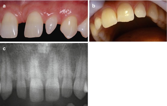
Conditions associated with the tooth size and arch length discrepancies which result in an imbalance between the width of the teeth and arch length are the most common causes of the diastema in adults [18, 19]. This can be due to microdontia, hypodontia, or increased arch dimensions. In other words, diastema occurs when the mesiodistal width of the anterior teeth is normal but the dental arch is larger (Fig. 2.5a, b) or the anterior teeth, particularly maxillary lateral incisors, are smaller than the normal size (peg-shaped) and the arch length is normal [7, 20] (Fig. 2.6a–c).


Fig. 2.4
Ugly duckling stage. (a, b) Obvious distal inclination of the maxillary incisors creating a midline diastema in the mixed dentition. (c) Maxillary incisors tend to upright by the eruption of the permanent canines and the midline diastema reduces. (d, e) Spontaneous closure of maxillary midline diastema after the canines are fully erupted

Fig. 2.5
(a, b) Generalized maxillary spacing due to tooth size–arch size discrepancy
It has been reported that small-sized or peg-shaped maxillary lateral incisors are the most common tooth size discrepancies among the tooth size abnormalities [4, 5]. This may result in localized spacing in the lateral incisor region or sometimes may lead to maxillary midline diastema due to distal migration of the central incisors into the space that have been formed at the mesial of the lateral incisors [5]. Similarly, when there is congenital or acquired absence of the lateral incisors, migration of the adjacent teeth creates generalized diastema [2, 5, 20] (Fig. 2.7a, b). Furthermore, the central incisors of the subjects with congenitally missing lateral incisors are likely to be smaller than the norms, leading to increased spacing. An association between hypodontia and microdontia has been revealed in the literature [21].


Fig. 2.6
(a) Peg-shaped maxillary lateral incisor. (b) Small-sized lateral incisor, (c) radiographic appearance of the figure b
2.1.4 Enlarged Labial Frenum and Deficient Intermaxillary Suture
Enlarged maxillary labial frenum has been reported as a cause or a consequence of the maxillary midline diastema [2, 5, 8] (Fig. 2.8a–c). At the beginning of 1900s, it has been described that the abnormal frenum was the cause of the maxillary midline diastema [22]. Shashua and Artun [8] have correlated existence of an enlarged labial frenum and occurrence of diastema in their study. During the eruption of the maxillary central incisors, a physiological space may exist between these teeth, and spontaneous closure of the space and atrophy of the labial frenum may occur at this stage after the eruption of the maxillary lateral incisors and canines [2, 23, 24]. However, sometimes this phenomenon does not occur, and the maxillary central incisors may erupt widely separated from each other. In this case, labial frenum can be attached deep within the tissue into the notch of the alveolar bone and create a heavily fibrous tissue between the central incisors causing a diastema [2, 23, 25]. In such cases, enlarged and deep fibrous frenum attachment does not change with age [5, 26] and will resist spontaneous closure of the space [3, 5, 12]. Contrary to these reports, it has been stated later that abnormal frenum was a consequence rather than the cause of diastema [2, 5, 27, 28]. It has been claimed that the eruption of the teeth, development of the alveolar process, and hypertrophic frenum occur simultaneously in the coronal direction. Thus, no or minimal pressure can be generated on the frenum to prevent the formation of diastema [28, 29]. It was previously reported that there were no correlation between the abnormal frenum and width of the diastema, width of frenum and diastema, or between the frenum length and width [2, 23]. Bergström et al. [27] concluded that the diastema closure progressed more rapidly in patients with surgically frenectomized group than in the non-frenectomized group, but the final results after 10 years were the same. In conclusion, enlarged or abnormal frenum can restrain the approximation of the maxillary central incisors, but may not be considered as an important etiological factor in maxillary midline diastema [2]. Before the management of this problem, a careful clinical evaluation should be carried on along with the patient’s age and other relevant parameters related to this condition [5, 24, 26
Stay updated, free dental videos. Join our Telegram channel

VIDEdental - Online dental courses


