When considering camouflage orthodontic treatment of a malocclusion associated with significant facial asymmetry, it is important to define the location of the dental midline. The patient, a 19-year-old Japanese woman, had an anterior open bite and a dental midline discrepancy associated with facial asymmetry. A nonsurgical treatment plan was considered. The main treatment objective was to correct the anterior open bite and the dental midlines in both arches. The dental midline discrepancy was eliminated, and proper overjet and overbite were achieved. Although the facial asymmetry remained, oral esthetics dramatically improved and a favorable occlusion was obtained. The results suggest that appropriately defining the location of the dental midline is critical for successful camouflage treatment of facial asymmetry.
Highlights
- •
Dental midline is critical for esthetics in camouflage treatment of facial asymmetry.
- •
For this patient, the dental midline was located on a smiling photograph.
- •
Oral esthetics were considerably improved by treatment.
- •
Good occlusion was obtained with favorable mesiodistal inclination of the incisors.
Facial asymmetry is one of the most challenging problems in orthodontics. Patients with severe facial asymmetry are generally treated with a combination of orthodontic and orthognathic surgical therapies to improve not only their occlusion, but also their facial esthetics. Although satisfactory results can be achieved by orthognathic surgery such as maxillomandibular osteotomy, in borderline cases, patients do not readily accept surgical orthodontics because of its invasiveness and accompanying risks. For these patients, we often perform camouflage orthodontic treatment. In camouflage treatment, it is important to define the location of the dental midline for oral esthetics. However, there is no standard to define the dental midline in camouflage treatment associated with facial asymmetry.
In this case report, we defined the location of the dental midline for oral esthetics in camouflage treatment by referring to a photograph of the patient smiling and obtained good results.
Diagnosis and etiology
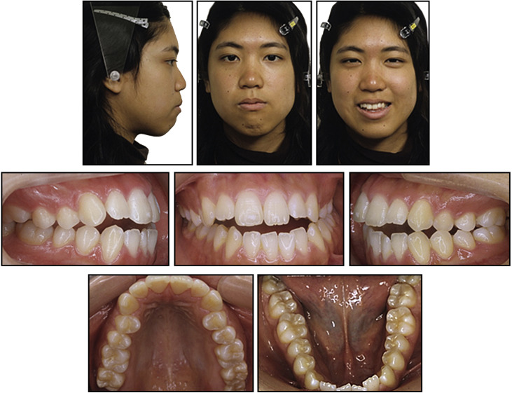
The patient was a 19-year-old Japanese woman. Her chief complaint was anterior open bite and discrepancy of the midlines between the maxillary and mandibular arches. She was not aware of her facial asymmetry and was not dissatisfied with or concerned about it. There was no history of trauma to her head or jaw. The family history was not relevant, and the cause of her facial asymmetry was unknown. In the frontal view, the mandible was slightly deviated to the left, and the lip line was canted. In the lateral view, the facial profile was straight, with upper and lower lip protrusion. The discrepancy between the maxillary and mandibular midlines was 4.0 mm, with the maxillary incisors inclined to the left and the mandibular incisors inclined to the right. Overbite and overjet were −2.0 and 4.0 mm, respectively. The occlusal plane was canted and almost parallel to the line passing through the left and right corners of the mouth. The anterior open bite extended from the left first premolar to the right first premolar. The molar relationships were Class III on the right and Class I on the left. There was no crossbite or scissorsbite in the posterior teeth, except in the right first premolar region ( Fig 1 ).
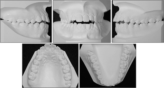
The dental casts showed transverse dental compensation of the incisors and the posterior teeth. The maxillary incisors were inclined to the left, and the mandibular incisors were inclined to the right. A clear midline discrepancy existed between the maxillary and mandibular arches. On the other hand, the maxillary posterior teeth were inclined to the left, and the mandibular posterior teeth were inclined to the right to maintain the buccolingual occlusion. There was minor crowding in the mandibular arch, and the mandibular arch length discrepancy was −2.1 mm ( Fig 2 ).
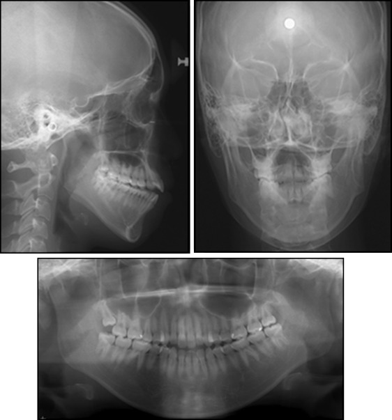
The lateral cephalometric analysis showed a skeletal Class I jaw relationship (ANB, 2.8°; Fig 3 ; Table ). The mandibular plane angle was steep. The maxillary central incisors were inclined labially. The frontal cephalometric analysis showed deformation of the maxilla and deviation of the mandible. The difference in height between the maxillary left and right molars was 1.8 mm, and the cant of the occlusal plane was 2.0°. Menton was deviated to the left by 5.4 mm from the facial midline.
| Variable | Norm (SD) | Pretreatment | Posttreatment | 1 year 6 months after treatment |
|---|---|---|---|---|
| Angular (°) | ||||
| SNA | 81.3 (3.0) | 78.5 | 78.7 | 78.7 |
| SNB | 79.2 (3.0) | 75.7 | 75.8 | 75.7 |
| ANB | 2.1 (2.1) | 2.8 | 2.9 | 3.0 |
| Facial angle | 87.3 (3.1) | 82.8 | 83.1 | 82.6 |
| FMA | 27.1 (5.2) | 37.1 | 36.5 | 37.0 |
| U1-SN | 104.3 (5.8) | 108.0 | 97.5 | 99.0 |
| FMIA | 58.0 (6.0) | 53.5 | 60.0 | 59.5 |
| IMPA | 93.0 (6.2) | 89.3 | 83.6 | 83.6 |
| Linear (mm) | ||||
| N-Me | 126.0 (4.7) | 142.8 | 142.2 | 142.9 |
| Ans-Me | 70.1 (4.4) | 85.2 | 84.6 | 85.3 |
| Is-PP | 29.8 (3.6) | 33.4 | 35.3 | 35.2 |
| U6-PP | 23.8 (2.5) | 27.4 | 27.0 | 27.5 |
| Ii-MP | 43.8 (1.9) | 51.4 | 52.1 | 52.0 |
| L6-MP | 34.2 (2.6) | 40.6 | 39.9 | 40.7 |
The final diagnosis was open bite and severe midline discrepancy associated with facial asymmetry.
Treatment objectives
The treatment objectives were as follows: (1) correct the facial asymmetry, (2) correct the anterior open bite, (3) correct the dental midlines in both arches, (4) obtain proper overjet and overbite, (5) correct the molar relationship, (6) establish a functional occlusion, and (7) improve lip protrusion.
Stay updated, free dental videos. Join our Telegram channel
VIDEdental - Online dental courses


