Crown Fracture with Pulp Exposure
OBJECTIVES
1 Recognize tissue changes resulting from pulp exposure and concomitant luxation injuries.
2 Determine treatment options.
3 Provide emergency and definitive care.
DESCRIPTION AND CLINICAL APPEARANCE
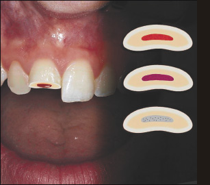
A fracture involving enamel and dentin with loss of tooth structure and exposure of the pulp (complicated crown fracture). Depending on the absence or presence of a concomitant luxation injury, the pulp will present with a bright red, cyanotic or ischemic appearance, respectively. There may be spontaneous bleeding from the pulp.
RADIOGRAPHIC APPEARANCE
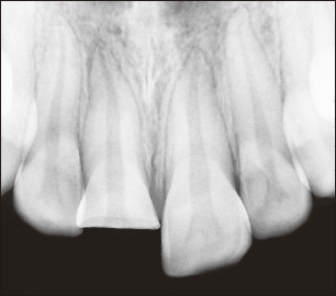
Lost tooth substance is apparent, as well as periodontal changes in the case of concomitant luxation.
BIOLOGICAL CONSIDERATIONS
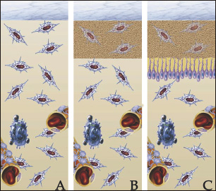
Exposed pulp tissue has a very good healing capacity in that it can normally close the perforation by forming a dentin barrier if a proper capping material is used.39,44–48 Clinical studies have shown that the length of exposure (days or weeks) does not reduce the chance of hard tissue healing. 47,48 Immediate or delayed treatment in the form of pulp capping or partial pulpotomy is the best solution when possible. This implies that the pulp can be left in the interim period before definitive treatment without a temporary coverage. Temporary coverage very often shows microleakage with penetration of anerobic bacteria into the exposed pulp, which represent a very harmful condition which may lead to an infected partial or total pulp necrosis. Proper capping or amputation m/>
Stay updated, free dental videos. Join our Telegram channel
VIDEdental - Online dental courses


