Introduction
Anchorage control is a challenge in orthodontics. Implants can be used to provide absolute anchorage.The aim of this study was to evaluate the success rates of palatal implants used for various anchorage purposes.
Methods
Thirty-four palatal implants were placed in 33 patients. In the adults (n = 9), the implants (n = 9) were placed in the median palatal suture. In the adolescents (n = 24), the implants (n = 25) were placed in the paramedian region. The implants were used to support a transpalatal arch, a modified distal jet appliance, or a modified hyrax screw. An implant was considered successful if it could be used as planned throughout the orthodontic treatment. The patients were asked to evaluate their pain perception after placement and explantation procedures.
Results
Three implants failed early (during the waiting period before orthodontic loading, within 3 months after placement). During the orthodontic loading period, no implants were lost. No statistically significant correlations were found between success rate and sex, age, primary stability, placement site (median or paramedian), implant size, or palatal depth. Pain perception after surgery was acceptable. The success rate of the palatal implants in this study was 91%.
Conclusions
Palatal implants are a reliable method of providing absolute anchorage control in a variety of patients for different indications. They can be loaded both directly and indirectly.
Anchorage control is a challenging problem in orthodontics. Several solutions have been proposed and tested. Grouping several teeth as anchorage has been suggested. Burstone and Kuhlberg tried to improve anchorage control by making use of the fact that tooth tipping is easier to achieve than axial or root movement. Extraoral anchorage has been suggested when anchorage in the dental arch is insufficient. None of these solutions has proven to be absolutely successful. For this reason, implant-like devices have been introduced in orthodontics for absolute anchorage. They are referred to as orthodontic implant anchors or temporary anchorage devices.
A placement site commonly used to provide anchorage with temporary anchorage devices is the palate, which has been studied by several investigators. The midpalatal suture area seems especially to be an ideal placement site, since it has both thin soft tissue and thick cortical bone. This is interesting when a screw is to be placed, since the main objective of an orthodontic screw is to gain maximum retention (good quality and quantity of bone) and prevent soft-tissue inflammation. Miniscrews can be placed nearly everywhere in the mouth and also in the midpalatal suture area.
One temporary anchorage device designed specifically to be used in this region is the palatal orthodontic implant anchor of the Orthosystem (Straumann, Basel, Switzerland). Although not as popular as miniscrews, palatal implants are gaining acceptance as an important alternative for achieving maximum intraoral orthodontic anchorage. In adult patients, the median palatal suture zone is the area of choice for placement of palatal implants. In adolescents, however, the paramedian region is preferred to avoid possible growth impairment of the maxilla in a transverse direction by placing an implant in the median palatal suture. The paramedian region has been described as a suitable placement site for implants. The site for palatal implants is standardized (median or paramedian palate); this is a major advantage, since a standard surgical procedure that is simple and easily controlled in all stages is essential for the success of an implant. Various rates of success for implants have been reported. For dental implants, 5-year cumulative success rates of 90% to 95% were reported ; success rates varied from 70% to 90% for miniscrews and from 84.8% to 100% for palatal implants.
The use of palatal implants is mainly indicated for adult patients. However, more adolescents are reluctant to wear extraoral appliances, with the result that compliance is greatly reduced with these treatment options. To avoid wearing extraoral appliances, children as young as 10 years now receive palatal implants.
The aims of this study were to evaluate (1) the success rate of palatal implants used for orthodontic purposes; (2) whether the success rate of palatal implants can be correlated to sex, age, primary stability, placement site (median or paramedian), form of the palate (wide or deep), implant size, and type of suprastructure (loading type); and (3) the patients’ pain after placement and removal.
The working hypothesis of this study was that a palatal orthodontic anchorage implant is a safe and predictable therapeutic technique, applicable to many clinical situations.
Material and methods
The study group consisted of 32 patients, each with 1 implant, and 1 patient with 2 implants. They were consecutive patients, treated at the Dental Clinic of the Department of Orthodontics at Vrije Universiteit Brussel in Jette, Belgium, between October 1998 and May 2003, who had finished their orthodontic treatment.
Table I shows the composition of the group by sex and age. A division was made between adolescent and adult patients. The adolescent group comprised 24 patients, all under the age of 16 years. The adult group comprised 9 patients, all above the age of 20 years.
| Adolescent girls | Adolescent boys | Women | Men | |
|---|---|---|---|---|
| Patients (n) | 11 | 13 | 8 | 1 |
| Mean age | 12 y 2 mo | 13 y 7 mo | 35 y 8 mo | 47 y 5 mo |
| Age range | 10 y 3 mo-14 y 6 mo | 11 y 5 mo-15 y 6 mo | 21 y 5 mo-53 y 2 mo | 47 y 5 mo |
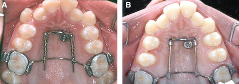
Most patients had an Angle Class II malocclusion. They were treated by extraction of the maxillary first or second premolars or distalization of the maxillary posterior segments. In the extraction patients, the implants were loaded indirectly and provided anchorage reinforcement for the posterior teeth (n = 18, Fig 1 ). When the aim of treatment was distalization of the maxillary posterior teeth, the implants were loaded directly and used to anchor a modified distal jet appliance for maxillary molar distalization (n = 13, Fig 2 ). In a patient with multiple agenesis, the implant was used to anchor a cantilever for aligning a palatally positioned canine. In 1 patient, 2 palatal implants were used to anchor a modified hyrax screw for skeletal palatal expansion (n = 2, Fig 3 ).


Stay updated, free dental videos. Join our Telegram channel

VIDEdental - Online dental courses


