Introduction
The purpose of this study was to determine the skeletal, dental, and soft-tissue changes in response to camouflage Class III treatment.
Methods
Thirty patients (average age, 12.4 ± 1.0 years) with skeletal Class III malocclusions who completed comprehensive nonextraction orthodontic treatment were studied. Skeletal, dental, and soft-tissue changes were determined by using published cephalometric analyses. The quality of orthodontic treatment was standardized by registering the peer assessment rating index on the pretreatment and posttreatment study models. The change in the level of gingival attachment with treatment was determined on the study casts. The results were compared with a group of untreated subjects. Data were analyzed with repeated measures analysis and paired t tests.
Results
The average change in the Wits appraisal was greater in the treated group (1.2 ± 0.1 mm) than in the control group (–0.5 ± 0.3 mm). The average peer assessment rating index score improved from 33.5 to 4.1. No significant differences were found for the level of gingival attachments between the treatment and control groups. The sagittal jaw relationship (ANB angle) did not improve with camouflage treatment. A wide range of tooth movements compensated for the skeletal changes in both groups. The upper and lower limits for incisal movement to compensate for Class III skeletal changes were 120° to the sella-nasion line and 80° to the mandibular plane, respectively. Greater increases in the angle of convexity were found in the treated group, indicating improved facial profiles. Greater increases in length of the upper lip were found in the treated group, corresponding to the changes in the hard tissues with treatment.
Conclusions
Significant dental and soft-tissue changes can be expected in young Class III patients treated with camouflage orthodontic tooth movement. A wide range of skeletal dysplasias can be camouflaged with tooth movement without deleterious effects to the periodontium. However, proper diagnosis and realistic treatment objectives are necessary to prevent undesirable sequelae.
A developing skeletal Class III malocclusion is one of the most challenging problems confronting an orthodontist. The prevalence of Class III malocclusion in the United States was approximately 1%. However, approximately 16% of patients aged 4 to 10 referred to an orthodontist have a diagnosis of Class III malocclusion.
Young patients who are diagnosed early with this problem can be treated orthopedically with a chincap or protraction facemask to normalize the underlying skeletal discrepancy. Patients with no growth remaining must be camouflaged by orthodontic tooth movement or fixed appliances. Camouflage treatment is the displacement of teeth relative to their supporting bone to compensate for an underlying jaw discrepancy. It implies that growth modification to overcome the basic problem is not feasible. The technique to camouflage a skeletal malocclusion was developed as an extraction treatment and introduced into orthodontics in the 1930s and 1940s. During that era, extraction to camouflage a skeletal malocclusion became popular because growth modification had been largely rejected as ineffective, and surgical correction had barely begun to develop. The strategy to camouflage a Class III malocclusion usually involves proclination of the maxillary incisors and retroclination of the mandibular incisors to improve the dental occlusion, but it might not correct the underlying skeletal problem or facial profile. Studies have shown an increase in the ANB angle, little or no change in the vertical dimension, and decreased concavity of the facial profile with Class III camouflage treatment. However, little information is available on possible tooth movements to camouflage this type of skeletal malocclusion. Our objective in this study was to determine the skeletal, dental, and soft-tissue changes in response to camouflage Class III treatment. The null hypothesis was that there are no significant differences in the skeletal, dental, and soft-tissue changes between treated and control Class III samples.
Material and methods
Forty-one patients, selected from the office files of an author (D.R.M.), had completed Class III camouflage treatment. The criteria for selection included (1) Class III molar relationship or mesial step in the mixed dentition, (2) concave facial profile, (3) Wits appraisal <–1.5 mm or ANB angle <1.0°, (4) a reduction in peer assessment rating (PAR) score >30%, (4) nonextraction comprehensive orthodontic treatment, and (5) high-quality pretreatment and posttreatment orthodontic records. Exclusion criteria included (1) dentofacial anomalies such as cleft lip and palate, (2) extracted or missing teeth, and (3) periodontal disease. Four patients were eliminated because they had extraction treatment; 5 were eliminated because of inadequate reductions in PAR scores; 2 were eliminated because there were no control subjects of similar age, sex, and craniofacial morphology to match them. No patient was eliminated because of poor records. The final sample consisted of 30 white patients (11 boys, 19 girls; average age, 12.4 ± 1.0 years). The mean treatment time was 2 years 2 months ± 7 months. Lateral cephalometric radiographs were taken before treatment (T1) and after treatment (T2). The cephalometric analyses used to evaluate skeletal, dental, and soft-tissue changes were described in the literature. The quality of orthodontic treatment was standardized by registering the PAR index on the pretreatment and posttreatment dental casts. Certified PAR calibration was obtained from Ohio State University, Columbus, Ohio, before this project. The change in the level of gingival attachment with treatment was measured on the study casts.
The control group consisted of serial radiographs of 30 white subjects (11 boys, 19 girls) from the Bolton-Brush Study in Cleveland, Ohio, who were matched by age, sex, and craniofacial morphology to the experimental sample. There were no significant differences in skeletal age between the treated and the control groups. A selection of cephalometric records describing the initial craniofacial morphology of the control and treated subjects is shown in Table I . Significant differences were found with variables OLp-A, molar relationship, SNA, SNB, ILG, Is/SNL, Is/FH, Ii/ML, U1-NL, and U1-L1.
| Skeletal and dental measurements | ||||||||
|---|---|---|---|---|---|---|---|---|
| Treated | Control | |||||||
| Variable | Mean | SD | Mean | SD | Difference | P value | Sig | |
| Sagittal (mm) | ||||||||
| Skeletal | Olp-A | 70.67 | 5.83 | 67.8 | 4.14 | 2.87 | 0.03 | ∗ |
| Olp-B | 75.69 | 7.87 | 72.72 | 5.83 | 2.97 | 0.1 | NS | |
| Olp-Pg | 79.69 | 9.09 | 76.20 | 6.45 | 3.49 | 0.09 | NS | |
| Wits | –7.16 | 2.81 | –6.14 | 2.31 | –1.02 | 0.12 | NS | |
| Co-ANS | 89.46 | 7.05 | 86.69 | 4.8 | 2.77 | 0.08 | NS | |
| Co-Pg | 113.34 | 9.37 | 109.45 | 6.46 | 3.89 | 0.06 | NS | |
| Dental | Is/Olp | 78.57 | 6.89 | 75.84 | 5.28 | 2.73 | 0.09 | NS |
| Ii/Olp | 76.4 | 6.48 | 73.90 | 5.64 | 2.5 | 0.11 | NS | |
| Overjet | 2.11 | 2.12 | 1.92 | 1.91 | 0.19 | 0.71 | NS | |
| Ms/Olp | 49.38 | 5.77 | 46.92 | 4.13 | 2.46 | 0.06 | NS | |
| Mi/Olp | 53.06 | 5.9 | 51.73 | 4.43 | 1.33 | 0.33 | NS | |
| Molar relationship | –3.7 | 2.01 | –4.82 | 1.94 | 1.12 | 0.03 | ∗ | |
| Vertical (mm) | ||||||||
| Skeletal | N-A | 50.22 | 4.68 | 48.27 | 3.18 | 1.95 | 0.06 | NS |
| ANS-Me | 63.58 | 6.43 | 62.2 | 4.76 | 1.38 | 0.34 | NS | |
| Dental | Is-NL | 26.71 | 2.82 | 26.14 | 2.56 | 0.57 | 0.41 | NS |
| Ii-ML | 36.68 | 4.36 | 35.96 | 2.56 | 0.72 | 0.43 | NS | |
| Overbite | 1.1 | 2.15 | 0.48 | 1.74 | 0.62 | 0.22 | NS | |
| Msc-NL | 21.49 | 2.54 | 20.93 | 2.31 | 0.56 | 0.37 | NS | |
| Mic-ML | 28.98 | 3.25 | 27.8 | 2.2 | 1.18 | 0.1 | NS | |
| ILG | 4.3 | 5.09 | 0.93 | 1.65 | 3.37 | 0.001 | † | |
| Angular (°) | ||||||||
| Skeletal | SNA | 79.56 | 3.54 | 74.32 | 3.79 | 5.24 | 0.0001 | † |
| SNB | 80.1 | 4.11 | 75.17 | 4.49 | 4.93 | 0.0001 | † | |
| ANB | –0.46 | 2.74 | –0.85 | 2.19 | 0.39 | 0.55 | NS | |
| ANL-ML | 33.68 | 6.16 | 32.97 | 5.74 | 0.71 | 0.64 | NS | |
| SNL-OL | 17.36 | 4.82 | 17.54 | 5.52 | –0.18 | 0.89 | NS | |
| SNL-NL | 7.66 | 3.75 | 7.52 | 3.45 | 0.14 | 0.87 | NS | |
| Dental | Is/SNL | 107.36 | 6.93 | 103.32 | 5.9 | 4.04 | 0.01 | ∗ |
| Is-FH | 118.03 | 6.77 | 114.36 | 4.53 | 3.67 | 0.01 | ∗ | |
| Ii/ML | 89.05 | 7.79 | 84.22 | 6.34 | 4.83 | 0.01 | ∗ | |
| U1-NL | 114.63 | 6.9 | 111.19 | 4.97 | 3.44 | 0.03 | ∗ | |
| U1-L1 | 129.91 | 10.61 | 119.7 | 7.33 | 10.21 | 0.0001 | † | |
The PAR index was used in this study to evaluate the quality of camouflage orthodontic treatment. The index was originally developed to assess how well orthodontic treatment reduces the severity of malocclusion. A score was assigned based on various occlusal traits that make up a malocclusion. The total score represents the degree to which a person’s occlusion deviates from normal alignment. The difference in scores between pretreatment and posttreatment reflects the improvement or success of the treatment. According to Feghali et al, a reduction in the PAR score of 22 or more points indicates “great improvement,” a reduction of 30% indicates an “improved condition,” and a reduction of less than 30% indicates “no improvement.” In this study, only patients who had a 30% or greater reduction in their PAR scores were included.
To assess the periodontium with the study casts, we measured the change in the level of gingival attachment with an electronic caliper (Ultra-Cal Mark III, Fowler-Sylvec, Boston, Mass) and a cephalometric protractor (3M Unitek, Monrovia, Calif) for the 4 mandibular incisors. Crown height was measured from the deepest point of the curvature of the facial vestibulogingival margin to the incisal edge of the incisors. Measurements were made to the nearest 0.1 mm with a Boley gauge. A paired t test was used to evaluate treatment changes (T2-T1) in the treated group and growth changes between the serial radiographs (t2-t1) in the control group. A repeated measures analysis was used to assess the differences between the treated and the control groups (T2-T1)-(t2-t1).
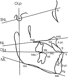
Cephalometric changes during treatment were evaluated on the lateral cephalometric radiographs taken at T1 and T2 for the treated sample and at t1 and t2 for the control sample. All radiographs were analyzed by using a combination of landmarks from various traditional cephalometric analyses ( Figs 1 and 2 ). Analysis of the sagittal skeletal and dental changes were recorded along the occlusal plane (OL) and to the occlusal plane perpendicular (OLp) obtained from the radiographs at t1 and T1. The OL and the OLp from the t1 and T1 tracings formed the reference grid for all sagittal measurements between OLp and the cephalometric landmarks. The grid was then transferred to the radiographs at t1 and T2, superimposing the tracings on the sella-nasion line (SNL) and along the anterior cranial base structure. The distance between OLp and the cephalometric landmarks were measured. Overjet and molar relationship were then calculated by summing the skeletal and dental contributions. A matched-pairs test was used to evaluate the significant treatment changes between T1 and T2 for the treated sample and t1 and t2 for the control sample. A repeated measures analysis was performed to evaluate the changes between the 2 samples (T2-T1)-(t2-t1).

Results
Errors in locating, superimposing, and measuring the changes of the landmarks were calculated. Cronbach’s correlation coefficient of reliability showed that all sagittal, vertical, and angular measurements and time periods were greater than 0.97; this indicates high reliability.
Sex differences were determined on all the tested variables. Significant differences were found only for the variable SN-NL and soft-tissue variables Ls-U1 and Ns-Ls/FH (data not shown). Male and female subjects were then combined for subsequent analyses. Table II shows the sagittal, vertical, and angular measurements at T1 and T2 for all subjects in the treated group. Significant differences were found in 10 of 12 variables in the sagittal measurements, 7 of 8 variables in the vertical measurements, and 1 angular measurement. Table III shows the sagittal, vertical, and angular measurements at t1 and t2 for all subjects in the control group. Significant differences were found in 10 of 12 variables in the sagittal measurements, 5 of 8 variables in the vertical measurements, and 6 angular measurements. Table IV compares the skeletal and dental changes between the treated and control groups. For sagittal changes, significant differences were found for the variables OLp-B, Wits, Is/OLp, and Ii/OLp. Greater forward movement of the mandible was found in the control group ( P <0.01). The Wits appraisal was decreased in the treatment group (–7.16 to –5.98) but increased in the control group (–6.14 to –6.67), P <0.002. The average maxillary incisor inclination was retroclined with treatment but proclined with growth in the control group ( P <0.02). The average mandibular incisor inclination was proclined with treatment but retroclined with growth in the control group ( P <0.03).
| Skeletal and dental measurements | ||||||||||||
|---|---|---|---|---|---|---|---|---|---|---|---|---|
| T1 | T2 | T2-T1 | ||||||||||
| Variable | Mean | SD | Min | Max | Mean | SD | Min | Max | Mean | P value | Sig | |
| Sagittal (mm) | ||||||||||||
| Skeletal | Olp-A | 70.67 | 5.83 | 59.9 | 83.5 | 72.48 | 6.24 | 61.3 | 88 | 1.81 | 0.0004 | ‡ |
| Olp-B | 75.69 | 7.87 | 60.8 | 92.3 | 77.63 | 8.53 | 62.6 | 95.5 | 1.94 | 0.0006 | ‡ | |
| Olp-Pg | 79.69 | 9.09 | 65 | 96.9 | 82.89 | 10.1 | 64.9 | 102.3 | 3.2 | 0.0001 | ‡ | |
| Wits | –7.16 | 2.81 | –12 | –1.5 | –5.98 | 2.92 | –12 | 1 | 1.18 | 0.002 | ‡ | |
| Co-ANS | 89.46 | 7.05 | 69.9 | 110.6 | 93.66 | 7.91 | 75 | 113.8 | 4.2 | 0.0001 | ‡ | |
| Co-Pg | 113.34 | 9.37 | 91.6 | 133.6 | 119.46 | 11.2 | 96.9 | 119.46 | 6.12 | 0.0001 | ‡ | |
| Dental | Is/Olp | 78.57 | 6.89 | 65.9 | 91.1 | 80.28 | 6.91 | 68.4 | 94.8 | 1.71 | 0.03 | ∗ |
| Ii/Olp | 76.4 | 6.48 | 54.1 | 87.1 | 78.38 | 7.2 | 66.6 | 92.1 | 1.98 | 0.002 | † | |
| Overjet | 2.11 | 2.12 | –5.5 | 6 | 2.03 | 1.28 | –2.6 | 3.8 | –0.08 | 0.53 | NS | |
| Ms/Olp | 49.38 | 5.77 | 37.1 | 60.2 | 53.02 | 5.31 | 43.4 | 63.8 | 3.64 | 0.0001 | ‡ | |
| Mi/Olp | 53.06 | 5.9 | 40.9 | 64 | 56.33 | 6.02 | 46.3 | 69.1 | 3.27 | 0.0001 | ‡ | |
| Molar relationship | –3.7 | 2.01 | –12.5 | –1.1 | –3.3 | 1.57 | –6.1 | 2.2 | 0.4 | 0.42 | NS | |
| Vertical (mm) | ||||||||||||
| Skeletal | N-A | 50.22 | 4.68 | 42.9 | 63.1 | 52.57 | 4.61 | 43.6 | 66.2 | 2.35 | 0.0001 | ‡ |
| ANS-Me | 63.58 | 6.43 | 53.1 | 74.1 | 67.4 | 6.9 | 55.4 | 77.8 | 3.82 | 0.0001 | ‡ | |
| Dental | Is-NL | 26.71 | 2.82 | 20.6 | 31.6 | 27.66 | 3 | 22.7 | 32.8 | 0.95 | 0.002 | † |
| Ii-ML | 36.68 | 4.36 | 23.1 | 42.7 | 39.27 | 3.64 | 33 | 45.4 | 2.59 | 0.0001 | ‡ | |
| Overbite | 1.1 | 2.15 | –5.2 | 5.3 | 1.04 | 0.87 | –0.8 | 3 | –0.06 | 0.85 | NS | |
| Msc-NL | 21.49 | 2.54 | 15.4 | 26.1 | 23.5 | 2.45 | 18.9 | 28.8 | 2.01 | 0.0001 | ‡ | |
| Mic-ML | 28.98 | 3.25 | 22.7 | 36.4 | 30.88 | 2.74 | 25.5 | 36.6 | 1.9 | 0.001 | ‡ | |
| ILG | 3.61 | 3.67 | 0 | 13.9 | 0.41 | 1.47 | 0 | 7.4 | –3.2 | 0.0003 | ‡ | |
| Angular (°) | ||||||||||||
| Skeletal | SNA | 79.56 | 3.54 | 70 | 87 | 78.38 | 4.23 | 70 | 89.0 | –1.18 | 0.32 | NS |
| SNB | 80.1 | 4.11 | 69.5 | 89 | 79.33 | 4.69 | 67.5 | 90 | –0.77 | 0.06 | NS | |
| ANB | –0.46 | 2.74 | –7 | 4 | –1.26 | 2.09 | –7 | 2 | –0.8 | 0.04 | ∗ | |
| ANL-ML | 33.68 | 6.16 | 21 | 53 | 33 | 7.55 | 20.5 | 58 | –0.68 | 0.69 | NS | |
| SNL-OL | 17.36 | 4.82 | 10 | 28 | 16.68 | 5.49 | 8 | 32 | –0.68 | 0.3 | NS | |
| SNL-NL | 7.66 | 3.75 | 0 | 14 | 8.08 | 4.47 | –2 | 18 | 0.42 | 0.24 | NS | |
| Dental | Is/SNL | 107.36 | 6.93 | 91 | 123 | 108.91 | 6.77 | 96.5 | 127 | 1.55 | 0.14 | NS |
| Is-FH | 118.03 | 6.77 | 108 | 138 | 120.1 | 6.91 | 109 | 138 | 2.07 | 0.07 | NS | |
| Ii/ML | 89.05 | 7.79 | 74 | 105 | 89.76 | 8.54 | 74 | 106 | 0.71 | 0.48 | NS | |
| U1-NL | 114.63 | 6.9 | 99 | 134 | 116.31 | 6.76 | 107 | 134 | 1.68 | 0.15 | NS | |
| U1-L1 | 129.91 | 10.61 | 109 | 148 | 128.2 | 10.09 | 108 | 144 | –1.71 | 0.28 | NS | |
| Skeletal and dental measurements | ||||||||||||
|---|---|---|---|---|---|---|---|---|---|---|---|---|
| t1 | t2 | t2-t1 | ||||||||||
| Variable | Mean | SD | Min | Max | Mean | SD | Min | Max | Mean | P value | Sig | |
| Sagittal (mm) | ||||||||||||
| Skeletal | Olp-A | 67.8 | 4.14 | 60.2 | 77.8 | 70.52 | 5.3 | 60.9 | 83.5 | 2.72 | 0.0001 | ‡ |
| Olp-B | 72.72 | 5.83 | 62.2 | 85.2 | 76.7 | 6.92 | 61.7 | 89.8 | 3.98 | 0.0001 | ‡ | |
| Olp-Pg | 76.2 | 6.45 | 64.6 | 89.5 | 81.04 | 7.67 | 66.1 | 94.2 | 4.84 | 0.0001 | ‡ | |
| Wits | –6.14 | 2.31 | –12.3 | –2.8 | –6.67 | 2.68 | –12.7 | –1.4 | –0.53 | 0.2 | NS | |
| Co-ANS | 86.69 | 4.8 | 77.3 | 95.4 | 90.99 | 4.86 | 83.9 | 102.3 | 4.3 | 0.0001 | ‡ | |
| Co-Pg | 109.45 | 6.46 | 97.5 | 123.8 | 116.38 | 6.43 | 106.3 | 131.7 | 6.93 | 0.0001 | ‡ | |
| Dental | Is/Olp | 75.84 | 5.28 | 67.2 | 88.5 | 79.14 | 6.36 | 66.7 | 92.6 | 3.3 | 0.0001 | ‡ |
| Ii/Olp | 73.9 | 5.64 | 62.6 | 86.7 | 77.72 | 6.56 | 66.7 | 93.3 | 3.82 | 0.0001 | ‡ | |
| Overjet | 1.92 | 1.91 | –2.5 | 8.3 | 1.39 | 2.34 | –3.8 | 8 | –0.53 | 0.03 | ∗ | |
| Ms/Olp | 46.92 | 4.13 | 39.6 | 57.1 | 50.55 | 6.13 | 39.8 | 65.5 | 3.63 | 0.0001 | ‡ | |
| Mi/Olp | 51.73 | 4.43 | 42.9 | 60 | 55.65 | 6.27 | 46.4 | 70.7 | 3.92 | 0.0001 | ‡ | |
| Molar relationship | –4.82 | 1.94 | –9.2 | –2 | –5.08 | 2.05 | –10.2 | –2.3 | –0.26 | 0.49 | NS | |
| Vertical (mm) | ||||||||||||
| Skeletal | N-A | 48.27 | 3.18 | 43.4 | 54.6 | 51.07 | 3.95 | 44.9 | 60.1 | 2.8 | 0.22 | NS |
| ANS-Me | 62.2 | 4.76 | 50.7 | 70.6 | 66.16 | 6.2 | 55.3 | 80.3 | 3.96 | 0.0001 | ‡ | |
| Dental | Is-NL | 26.14 | 2.56 | 19.3 | 30.5 | 26.84 | 2.89 | 20.5 | 32.2 | 0.7 | 0.007 | † |
| Ii-ML | 35.96 | 2.56 | 28.2 | 42.1 | 38.35 | 3.28 | 32.2 | 46.9 | 2.39 | 0.0001 | ‡ | |
| Overbite | 0.48 | 1.74 | –3.9 | 6.5 | 0.22 | 0.86 | –1.3 | 1.9 | –0.26 | 0.43 | NS | |
| Msc-NL | 20.93 | 2.31 | 14.9 | 25.1 | 23.09 | 2.57 | 17.8 | 28.1 | 2.16 | 0.0001 | ‡ | |
| Mic-ML | 27.8 | 2.2 | 23.6 | 32.8 | 29.95 | 3.2 | 24.5 | 38.5 | 2.15 | 0.0001 | ‡ | |
| ILG | 0.93 | 1.65 | 0 | 6.7 | 0.62 | 1.2 | 0 | 4.6 | –0.31 | 0.4 | NS | |
| Angular (°) | ||||||||||||
| Skeletal | SNA | 74.32 | 3.79 | 64.2 | 82.1 | 74.96 | 3.45 | 65.6 | 81.2 | 0.64 | 0.07 | NS |
| SNB | 75.17 | 4.49 | 63.2 | 82.1 | 76.53 | 4.28 | 64.2 | 85 | 1.36 | 0.0001 | ‡ | |
| ANB | –0.85 | 2.19 | –8.5 | 3.8 | –1.56 | 2.33 | –7.6 | 2.8 | –0.71 | 0.01 | ∗ | |
| ANL-ML | 32.97 | 5.74 | 24.5 | 44.4 | 31.72 | 6.58 | 20.8 | 48.1 | –1.25 | 0.03 | ∗ | |
| SNL-OL | 17.54 | 5.52 | 8.5 | 34 | 15.2 | 4.27 | 8.5 | 22.7 | –2.34 | 0.001 | † | |
| SNL-NL | 7.52 | 3.45 | 1.4 | 14.2 | 7.87 | 3.79 | 0 | 16 | 0.35 | 0.33 | NS | |
| Dental | Is/SNL | 103.32 | 5.9 | 89.7 | 113.3 | 105.11 | 7.08 | 88.7 | 118 | 1.79 | 0.04 | ∗ |
| Is-FH | 114.36 | 4.53 | 105.7 | 122.7 | 116 | 6.65 | 99.1 | 131.2 | 1.64 | 0.08 | NS | |
| Ii/ML | 84.22 | 6.34 | 70.8 | 93.5 | 83.12 | 5.94 | 69.9 | 93.5 | –1.1 | 0.1 | NS | |
| U1-NL | 111.19 | 4.97 | 97.2 | 122.7 | 113.07 | 6.03 | 99.1 | 129.3 | 1.88 | 0.03 | ∗ | |
| U1-L1 | 119.7 | 7.33 | 107.6 | 136.9 | 120.17 | 8.06 | 104.8 | 135.9 | 0.47 | 0.7 | NS | |
| Skeletal and dental measurements | |||
|---|---|---|---|
| Variable | P value | Sig | |
| Sagittal measurements (mm) | |||
| Skeletal | Olp-A | 0.13 | NS |
| Olp-B | 0.01 | † | |
| Olp-Pg | 0.06 | NS | |
| Wits | 0.002 | † | |
| Co-ANS | 0.92 | NS | |
| Co-Pg | 0.57 | NS | |
| Dental | Is/Olp | 0.02 | ∗ |
| Ii/Olp | 0.03 | ∗ | |
| Overjet | 0.29 | NS | |
| Ms/Olp | 0.99 | NS | |
| Mi/Olp | 0.48 | NS | |
| Molar relationship | 0.28 | NS | |
| Vertical measurements | |||
| Skeletal | N-A | 0.29 | NS |
| ANS-Me | 0.86 | NS | |
| Is-NL | 0.5 | NS | |
| Ii-ML | 0.72 | NS | |
| Overbite | 0.67 | NS | |
| Dental | Msc-NL | 0.74 | NS |
| Mic-ML | 0.69 | NS | |
| ILG | 0.0009 | ‡ | |
| Angular measurements | |||
| Skeletal | SNA | 0.32 | NS |
| SNB | 0.0001 | ‡ | |
| ANB | 0.85 | NS | |
| SNL-ML | 0.07 | NS | |
| SNL-OL | 0.08 | NS | |
| SNL-NL | 0.88 | NS | |
| Dental | Is/SNL | 0.85 | NS |
| Is-FH | 0.76 | NS | |
| Ii/ML | 0.14 | NS | |
| U1-NL | 0.89 | NS | |
| U1-L1 | 0.27 | NS | |
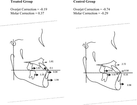
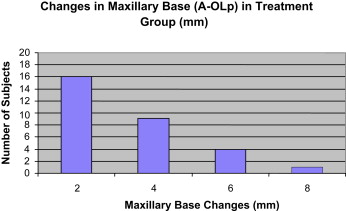
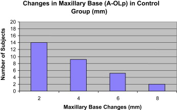
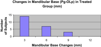
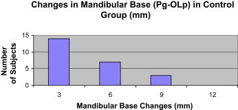
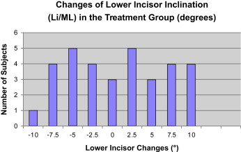
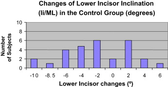
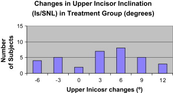
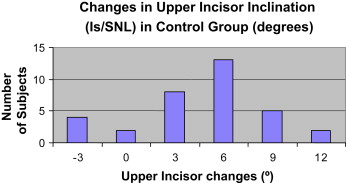
No significant differences were found in overjet between the treated and control groups ( Fig 3 ). The average changes in overjet in the treated and control groups were –0.19 and –0.74 mm, respectively. However, large variations in skeletal and dental changes were found in both groups that contributed to the changes in overjet ( Figs 4-11 ). The changes ranged from 2 to 8 mm in the maxillary base and 3 to 9 mm in the mandibular base in both groups. A similar distribution was found in both groups. A wide range of incisal changes was also noted in both groups to compensate for the skeletal changes. The changes in mandibular incisor inclinations ranged from –10° to 15° in the treated group and –10° to 6° in the control group. The changes in maxillary incisor inclinations ranged from –6° to 12° in the treated group and –3° to 12° in the control group. No significant differences were found in molar relationships between the 2 groups. The average changes in molar relationship for the treated and control groups were 0.37 and –0.27 mm, respectively.
For vertical changes, significant differences were found between the treated and the control groups for interlabial distance (ILG). A greater decrease in ILG was found in the treated group.
For angular changes, significant differences between the treated and control groups were found for the variable SNB. The forward movement of the mandibles (SNB) was less in the treated group compared with the control group.
Table V shows the sagittal, vertical, and angular soft-tissue measurements at T1 and T2 for all subjects in the treated group. Significant differences were found in 2 of 11 sagittal soft-tissue profile variables, 5 of 6 variables in vertical measurements, and 2 of 6 variables in soft-tissue thickness measurements. Table VI shows the sagittal, vertical, and angular soft-tissue measurements at t1 and t2 for all subjects in the control group. Significant differences were found in 4 of 11 variables in soft-tissue thickness measurements, 5 of 6 variables in vertical measurements, and 4 of 6 variables in soft-tissue thickness measurements. No significant differences were found in the lip-structure measurements. Table VII compares the soft-tissue changes between the treated and control groups. For soft-tissue profile changes, significant differences were found for the variables Ns-Sls/SLs-Pos, Ls/Pn-Pos, Ns-St, Ns-Li, and Ns-Pog. A greater increase in facial convexity was found in the treated group compared with the control group. For vertical changes, significant differences were found for the variable Sn-St. A greater increase in upper lip length was found in the treated compared with the control group.
| Total soft-tissue measurements | |||||||||||
|---|---|---|---|---|---|---|---|---|---|---|---|
| T1 | T2 | T2-T1 | |||||||||
| Mean | SD | Min | Max | Mean | SD | Min | Max | Mean | P value | Sig | |
| Sagittal relationship of soft tissue profile | |||||||||||
| Ns-Sls/ | –4.89 | 8.05 | –22 | 15.6 | –7.12 | 6.3 | –20.6 | 9 | –2.23 | 0.09 | NS |
| Sls-Pos(mm) | |||||||||||
| Ls/Pn-Pos (mm) | 81.67 | 10.21 | 62.6 | 100.4 | 81.74 | 10.55 | 62.6 | 105.9 | 0.07 | 0.93 | NS |
| Li/Pn-Pos (mm) | 80.08 | 15.47 | 15.5 | 103.6 | 82.76 | 11 | 63.4 | 106.5 | 2.68 | 0.37 | NS |
| Pn/Ns (mm) | 26.56 | 4.77 | 17.8 | 38 | 28.08 | 5.24 | 20.3 | 39.7 | 1.52 | 0.0005 | ‡ |
| Ns-Sn (mm) | –12.41 | 6.77 | –25.5 | 11.7 | –11.33 | 14.73 | –24.5 | 65 | 1.08 | 0.54 | NS |
| Ns/Sls (mm) | –11.41 | 6.77 | –25.7 | 11.9 | –11.87 | 7.03 | –24.2 | 15.7 | –0.46 | 0.32 | NS |
| Ns/Ls (mm) | –16.04 | 8.4 | –31.8 | 18.3 | –15.44 | 6.84 | –29.8 | 7.6 | 0.6 | 0.33 | NS |
| Ns-St (mm) | –11.06 | 6.53 | –26.3 | 11.1 | –10.07 | 6.09 | –25.1 | 1 | 0.99 | 0.17 | NS |
| Ns/Li (mm) | –17.21 | 8.36 | –35.3 | 15.8 | –16 | 8.63 | –33.7 | 18.8 | 1.21 | 0.02 | ∗ |
| Ns/Ils (mm) | –12.27 | 7.58 | –31.4 | 16 | –12.03 | 6.55 | –30.4 | 1.8 | 0.24 | 0.72 | NS |
| Ns-Pog (mm) | –17.31 | 8.01 | –38.5 | 8.5 | –17.35 | 8.61 | –39.3 | 9.7 | –0.04 | 0.93 | NS |
| Vertical relationship of soft tissue profile | |||||||||||
| Sn-Ms (mm) | 69.68 | 6.54 | 60 | 82.2 | 72.89 | 6.43 | 61 | 84 | 3.21 | 0.0001 | ‡ |
| Sn-St (mm) | 18.55 | 2.83 | 12.9 | 23.4 | 21.59 | 2.53 | 13.7 | 25.7 | 3.04 | 0.0001 | ‡ |
| St-Ms (mm) | 47.73 | 5.28 | 37.8 | 58.2 | 50.52 | 5.33 | 39.8 | 60.9 | 2.79 | 0.0001 | ‡ |
| St-Ils (mm) | 16.88 | 2.47 | 12.1 | 21.5 | 17.4 | 2.34 | 12.1 | 22.1 | 0.52 | 0.18 | NS |
| Ns-Ms (mm) | 120.23 | 10.11 | 103.2 | 142.4 | 125.03 | 10.31 | 105.4 | 145.3 | 4.8 | 0.0001 | ‡ |
| Ns-Sn (mm) | 53.2 | 5.68 | 43.2 | 66.5 | 55.26 | 5.86 | 45.8 | 71.2 | 2.06 | 0.0001 | ‡ |
| Soft-tissue thickness | |||||||||||
| Sn-A (mm) | 15.81 | 2.95 | 9.9 | 23 | 17.33 | 2.79 | 10.2 | 23 | 1.52 | 0.006 | † |
| Ls-U1 (mm) | 13.27 | 2.51 | 8.9 | 19 | 13.36 | 2 | 9.7 | 17.2 | 0.09 | 0.81 | NS |
| Li-L1 (mm) | 14.3 | 2.37 | 9.1 | 19.5 | 14 | 2.29 | 10.7 | 22.7 | –0.3 | 0.41 | NS |
| Pos-Pog (mm) | 11.68 | 1.72 | 7.7 | 16.4 | 11.59 | 1.9 | 8.5 | 15.9 | –0.09 | 0.73 | NS |
| Sls-A (mm) | 14.99 | 3.1 | 8.1 | 21.9 | 16.25 | 2.55 | 9.1 | 21.2 | 1.26 | 0.01 | ∗ |
| Ils-B (mm) | 11.27 | 1.83 | 7.7 | 15.6 | 12.05 | 2.32 | 9 | 21.1 | 0.78 | 0.11 | NS |
| Lip structure | |||||||||||
| Ns-Ls/FH (°) | 100.51 | 4.89 | 93 | 115 | 99.34 | 5.57 | 77 | 108 | –1.17 | 0.31 | NS |
| Li-Ils/FH (°) | 52.65 | 14.44 | 17 | 81 | 53.1 | 11.24 | 24 | 71 | 0.45 | 0.84 | NS |
| Ils-Pos-Ls (°) | –17.6 | 9.69 | –32 | 17 | –17.65 | 5.12 | –26 | –10 | –0.05 | 0.97 | NS |
| Li/Pos-Ls (°) | 1.93 | 5.54 | –7 | 12 | 0.75 | 4.77 | –9 | 13 | –1.18 | 0.18 | NS |
Stay updated, free dental videos. Join our Telegram channel

VIDEdental - Online dental courses


