CEMENTUM
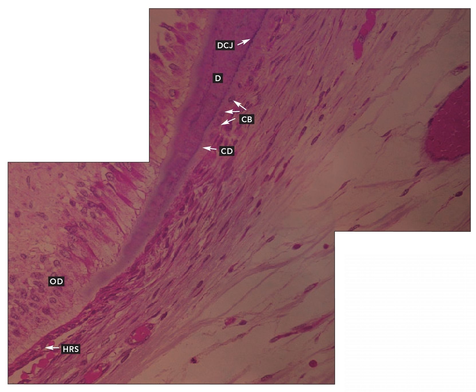
FIG 5-1
Cementum
Cementum forming on a developing root. Cementoblasts (CB) have deposited a layer of cementoid (CD) on the surface of root dentin (D) at the dentinocemental junction (DCJ). Hertwig’s root sheath (HRS) has induced more odontoblasts (OD) from the pulp (H and E stain; ×400).
Dentinocemental junction
Transverse section of a tooth in situ. Acellular cementum (AC) has been deposited on the dentin (D). The dentinocemental junction (DCJ) is prominent. Periodontal ligament (PDL) extends from the cementum to the alveolar bone (B) (H and E stain; ×160).
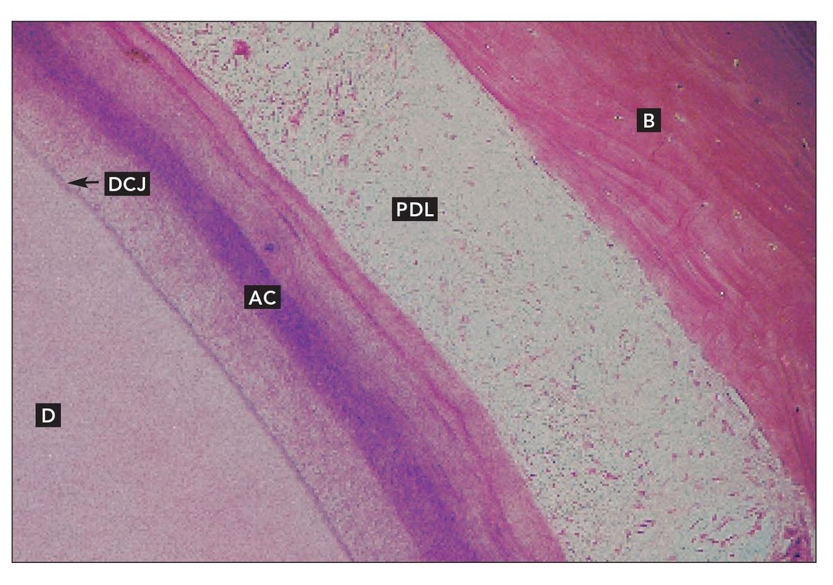
FIG 5-3
Dentinocemental junction
Higher magnification of the dentinocemental junction (DCJ), acellular cementum (AC), and periodontal ligament (PDL) shown in Fig 5-2 (×400).
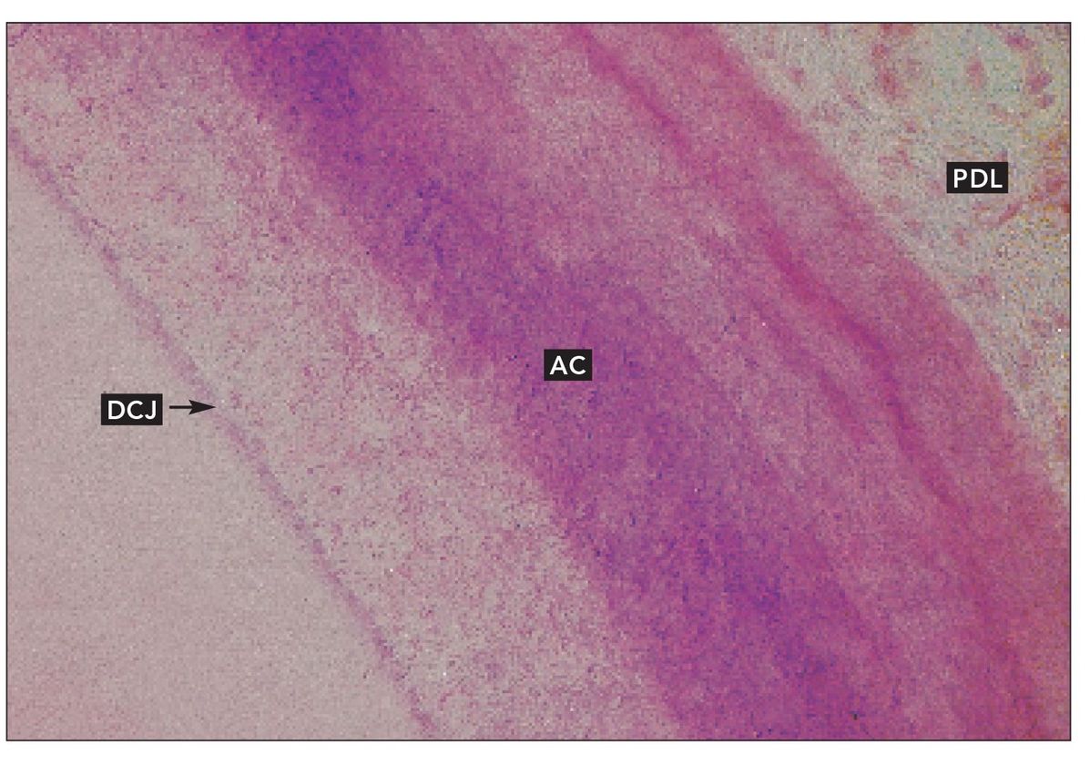
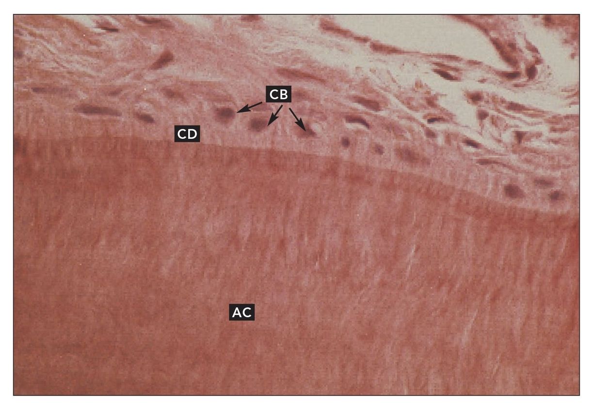
FIG 5-4
Acellular cementum
Cementoblasts (CB) and cementoid (CD) on the surface of acellular cementum (AC) (H and E stain; ×640).
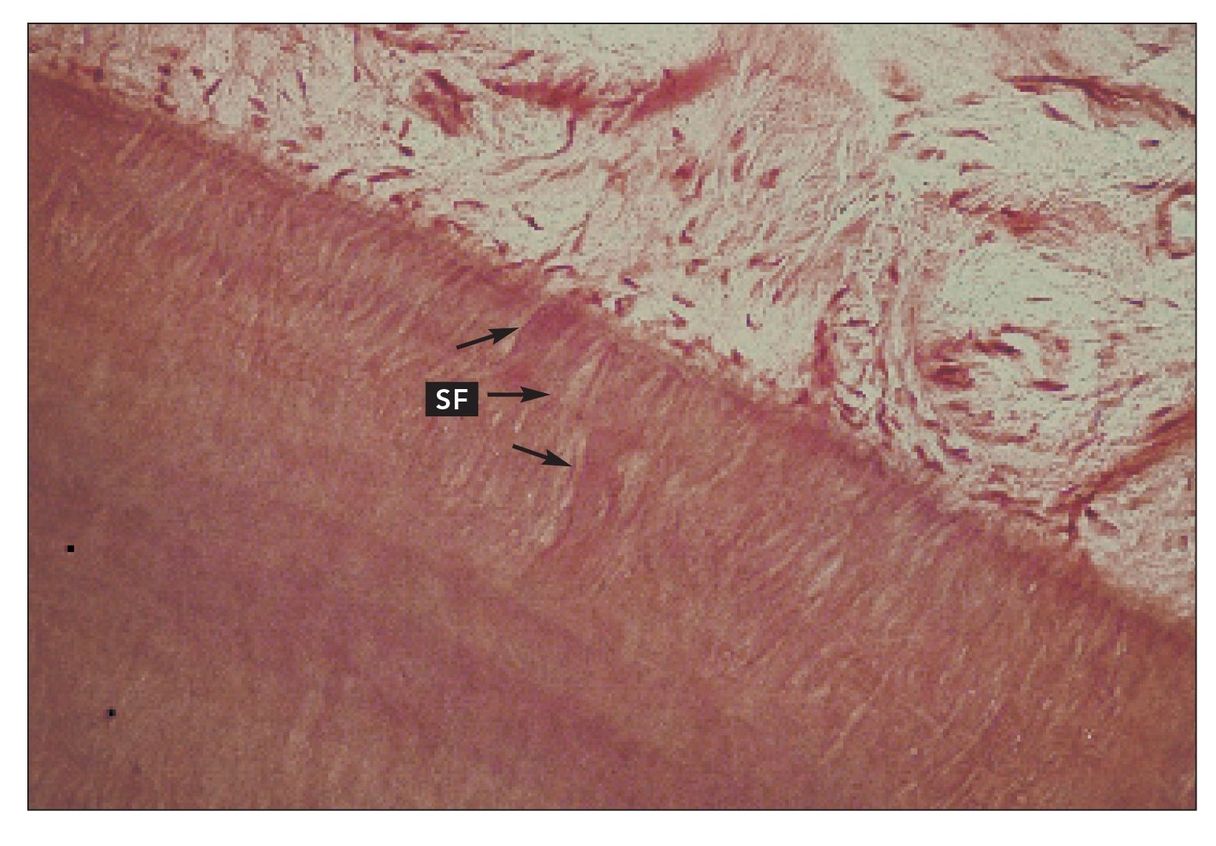
FIG 5-5
Sharpey’s fibers
Sharpey’s fibers (SF) in acellular cementum (H and E stain; ×400).
Stay updated, free dental videos. Join our Telegram channel
VIDEdental - Online dental courses


