and Jasdeep Kaur1
(1)
Earth and Life Sciences Vrije Universiteit Amsterdam and ILEWG, Amsterdam, The Netherlands
8.1 Introduction
Abstract
Bite mark evidence found on the skin of a living person or a dead body and on objects may be of great importance in criminal and legal investigations. This chapter focuses on digital bite mark analysis and the stepwise process of analysis.
8.1 Introduction
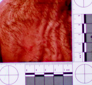
Bite mark (Fig. 8.1) evidence found on the skin of a living person or a dead body and on objects may be of great importance in criminal and legal investigations (Vale 1986). The method for comparing the bite marks is well recognized and includes measurement and analysis of the pattern, size, and shape of teeth against similar characteristics observed in an injury on skin or a mark left on an object (ABFO 1995). The most frequent analysis methods are used to produce life-sized comparison overlays from a suspect’s teeth to detect similarities or differences with the bite mark. Various methods exist to produce these overlays; in most techniques, the perimeter of the biting edges of the suspect’s teeth are hand-traced directly from dental study casts or from wax bite exemplars, or indirectly from xerographic images produced with office photocopiers that are calibrated to produce life-sized final images. But these methods cannot avoid the bias innate in observer subjectivity, and significant errors incorporated in the overlays may make it difficult to reach conclusions with a high degree of confidence in court and medical legal proceedings. Therefore, the AFBO has proposed an objective method using Adobe Photoshop® (ABFO 1995). Since biting is a forceful procedure involving three moving systems—the maxilla, the mandible and the victim’s reaction—most recent research has focused on analyzing how these factors affect the bite mark with the application of 3D procedures (Thali et al. 2003; Martin-de las Heras et al. 2005). The functional tools within Photoshop® can be used to detect and correct for certain angular distortions. This is an extremely important step, as it forms the foundation for the comparison procedures that follow (Johansen and Bowers 2003).


Fig. 8.1
Human bite marks
8.2 Advantage of the Digital Analysis of Bite Marks (Johansen and Bowers 2003)
Digital analysis of bite marks has a much faster processing speed, which saves time. Also, it is easy to distribute digital information. It is also easy to store a large number of images. Finally, it is a more objective method, with minimal bias.
8.3 Ideal Physical Evidence (Johansen and Bowers 2003)
As bite marks are photographed or dental radiographs are used as evidence, forensic odontologists should attempt to obtain a precise image of the bite marks. Thus, a tripod should be used even though off-angle distortion is really impossible to avoid. Ideally, photos of bite marks of two- and three-dimensional evidence should have the following criteria:
1.
ABFO No. 2 scale and bite marks should be in the same plane.
2.
The direction of the camera and the scale should be parallel.
3.
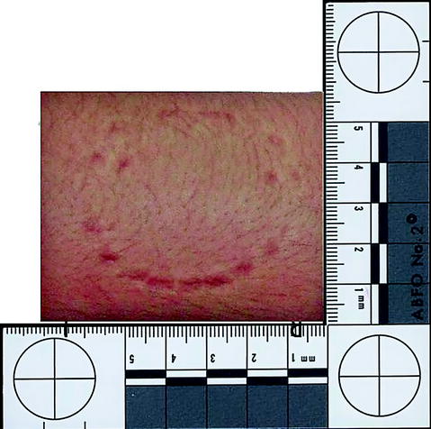
The bite mark should be photographed perpendicular to the ABFO No. 2 scale (Fig. 8.2).


Fig. 8.2
Original image of bite marks
4.
Avoid creating a parallax distortion of the scale caused by placing the scale away from the plane of the bite mark.
8.4 Stepwise Bite Mark Analysis by Taking Digital Images
The following steps explain how to use Adobe Photoshop to take a digital image of a bite mark:
1.
Import the image of the bite mark by clicking File; then click Open and open the Images file (Adobe Photoshop CS5®).
2.
Click on File, and save images as PSP.
3.
Prepare bite mark image by using the measure tool and rotation tool (click measurement tool and draw a line along one leg of the ABFO No. 2 scale).
4.
A rotating angle appears.
Stay updated, free dental videos. Join our Telegram channel

VIDEdental - Online dental courses


