Fig. 6.1
A polarized light microscope image of an erosive tooth wear with a crater formed by tooth substance loss (x), a layer of softened tooth tissue (y) and the sound enamel (z). X150
6.2 Objective and Quantitative Monitoring of Erosion
6.2.1 Silicone Matrix
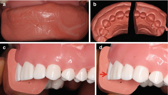
A simple and more reliable tool for monitoring the progression status of an erosive lesion over time is the silicone matrix system described by Shaw et al. [3]. This is an objective and quantitative clinical method of monitoring erosive tooth wear. It is one of the easiest and most useful methods of monitoring tooth wear. This is no difference from the simple method used by dental students to measure and monitor tooth tissue reduction during crown preparation. With this system, a silicone putty impression of the teeth is taken in an “unglued” sectional tray (Fig. 6.2a). The putty is removed from the tray, and it is sliced into sections through the centers of the erosive lesions (Fig. 6.2b). When a section is replaced on tooth surface, it is a perfect fit to the tooth surface (Fig. 6.2c). If the erosive lesion progresses, on the next review visit a gap will become visible between the silicone and the lesion surface (Fig. 6.2d). The depth of the gap can be measured in millimeters (mm) using the calibrated periodontal probe (explorer). An erosive wear can be confirmed arrested when there is no gap after several review visits. Besides being objectively quantitative, the silicone matrix enables slight change in depth and extent of the lesion to be detected and measured, which is its major advantage over the other methods discussed below. Generating quantitative data would also permit quantitative comparison of lesions at different time points, and possible statistical comparative analysis of the erosion progression over time.


Fig. 6.2
An illustration of the procedure for using silicone matrix to monitor erosive lesion progression status over time. (a) Silicone putty impression of the teeth is taken in an “unglued” sectional tray. (b) The putty is removed from the tray, and it is sliced into sections through the center of the erosive lesion. (c) When a section is replaced on tooth surface, it is a perfect fit to the tooth surface. (d) If the erosive lesion progresses, a gap (red arrowed) will become visible between the silicone and the lesion surface
6.2.2 Other Emerging Quantitative Systems
Following the demonstration of the feasibility of using the ultrasound system to measure enamel thickness [4], measurement of enamel thickness using a high-frequency ultrasonic transducer-based hand-held probe was used to follow the progression of enamel erosion by acid dissolution [5]. However, a study showed that due to measurement variation, thickness changes of less than 0.33 mm cannot be detected reliably [6]. Besides, ultrasound systems can only quantify tissue loss limited to enamel. Wilder-Smith et al. [7] used optical coherence tomography (OCT) to successfully monitor the progression status of erosive tooth wear in patients with GERD; however, just like the ultrasound system, OCT can only measure tissue loss limited to enamel.
6.3 Subjective and Semi-quantitative Monitoring of Erosion
6.3.1 Diagnostic Indices
The diagnostic indices score the severity of erosive lesions to mirror the condition in an index value, hence its semi-quantitative nature. The severity of an erosive lesion is scored by estimating the percentage of the entire surface affected by the condition and by whether dentin is exposed or not. The diagnostic indices are subjective evaluations, and are faced with the difficulty in distinguishing between lesions restricted to enamel and lesions with exposed dentin because exposed dentin cannot be reliably diagnosed by clinical means [8]. Besides, dentin exposure may not necessarily relate to extensive tissue loss, particularly in areas where the enamel covering is thin, such as in cervical regions of the teeth. Unlike the silicone matrix, diagnostic indices also have the problem of inability to detect slight change in lesion severity, particularly if the progression is in depth and not in area of the lesion. The following indices have been proposed [9], but none has been generally accepted.
6.3.1.1 Basic Erosive Wear Examination
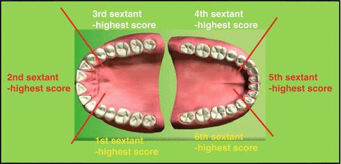
Most of the available indices were designed for epidemiological studies rather than for individual patients management in general practice and were not related to treatment needs. This has led to the proposal of a management-based index, the Basic Erosive Wear Examination (BEWE), by Bartlett et al. [10]. With the BEWE, the severity level of a lesion is scored on a four grade level based on its extent on the tooth surface (Table 6.1). The dentition is divided into sextants (Fig. 6.3), and the buccal, occlusal and lingual surfaces of every tooth in each sextant is examined for erosive wear and awarded a score value between 0 and 3. Then the most severely affected surface in a sextant is recorded. The sum of the scores from the sextants constitutes the index value (BEWE score), and guides the recommendations for the management of the condition by the practitioner (Table 6.2). The BEWE can be used to determine the erosion risk status of an individual patient [11]. Also the BEWE is a useful tool for monitoring the progression status of erosive lesions. However, as stated above, the subjective nature of the BEWE makes it difficult, if not impossible, for slight progression of the erosive lesion to be detected. Furthermore, with the BEWE, there is the possibility of ignoring newly developed erosive lesions.

Table 6.1
Criteria for grading erosive tooth wear by the Basic Erosive Wear Examination [10]
|
Scores
|
Erosive wear severity levels
|
|---|---|
|
0
|
No erosive tooth wear
|
|
1
|
Initial loss of surface texture
|
|
2a
|
Distinct defect, hard tissue loss <50 % of the surface area
|
|
3a
|
Hard tissue loss ≥50 % of the surface area
|

Fig. 6.3
An illustration of the division of the dentition into sextants for the Basic Erosive Wear Examination (BEWE)
Stay updated, free dental videos. Join our Telegram channel

VIDEdental - Online dental courses


