Introduction
Our aim was to evaluate the risk of external apical root resorption (EARR) in mesialized mandibular molars due to space closure in patients with unilateral second premolar agenesis. The contralateral side served as the control.
Methods
After application of eligibility criteria, 25 retrospectively selected subjects (median age, 14.9 years; range, 12.0-31.9 years) were analyzed. Space closure (approximately 10 mm) was performed using skeletal anchorage. EARR was measured at the mandibular permanent canines, first premolars, and first molars in the pretreatment and posttreatment orthopantomograms. Measurements were performed by 2 examiners independently and were corrected for distortion and magnification of radiographs, which were assessed in a pilot study. Multivariate analysis of covariance and pairwise comparisons were performed.
Results
The mean enlargement factor of the panoramic machine was 29% ± 0.3%. Distortion exceeded 5% only in cases of large positioning errors (>20°). Intraclass correlation coefficients showed strong to almost perfect agreement (mean, 0.80 mm; 95% CI, 0.75-0.85) of the two examiners. Multivariate analysis of covariance resulted in no difference in EARR between the canines and premolars of the space closure and control sides. On the contrary, there was a statistically significant difference between mesialized and nonmezialized molars (0.73 mm; 95% confidence interval, 0.19-1.27). The mean total EARR in each tooth type did not exceed 1 mm.
Conclusions
Space closure through extensive tooth movement in the mandible was identified as a risk factor for EARR. However, the amount of EARR attributed to space closure and the total EARR were not considered clinically significant.
Highlights
- •
Space closure with extensive tooth movement is clearly a risk factor for EARR.
- •
The amount of EARR attributed to space closure was not clinically significant.
- •
In terms of EARR, space closure through mesialization is a safe treatment option.
External apical root resorption (EARR) is an inevitable adverse effect of orthodontic treatment. Several studies have shown that the maxillary incisors usually show more EARR than any other teeth, followed by the mandibular incisors and first molars. In most patients, root shortening during treatment is limited to 2 mm or less. However, 1% to 5% of all orthodontic patients experience severe resorption, defined as more than 4 mm or a third of root shortening. Although in most cases the extent of EARR is limited and does not affect tooth survival, it is important for the practitioner to know which risk factors predispose the patient to the development of EARR to be able to manage this adverse effect of treatment. This is especially true when considering that severe chronic periodontal problems coincide with increased root resorption. Thus, the combined EARR and periodontal problems may result in severe loss of periodontal support, including an increased crown-root ratio, which may question long-term tooth viability.
The amount of EARR in a patient is mostly unpredictable before treatment. Recent studies have suggested various risk factors that could contribute to the development of EARR in a specific patient. Genetic predisposition is considered a major factor in determining EARR potential during orthodontic treatment. Additional patient-related risk factors include previous history of EARR, as well as type and severity of malocclusion to be treated. The orthodontic treatment-related risk factors include increased treatment duration, increased magnitude of applied force, tooth extraction treatment, intrusive movement, and increased tooth movement.
Prediction studies based on multivariate models that included certain risk factors for EARR failed to explain a considerable amount of the observed variation (explained variance, approximately 20%). Among others, this indicates unidentified risk factors. The amount of explained variance considerably increased (approximately 70%) when individual susceptibility was incorporated in the model through the amount of EARR that occurred during the first stages of orthodontic treatment. Thus, because of the multifactorial etiology of this phenomenon and the variability in study protocols that investigated specific risk factors for EARR development, the pretreatment prediction of EARR in a specific patient still remains impossible. Split-mouth study designs could be a valid alternative to control for individual susceptibility when testing the involvement of a specific risk factor in EARR development, but such studies are not available so far.
Various methods have been used to assess the amount of EARR in a patient. Certain studies have used specific drawings as a graded scale for classifying EARR, whereas others used length measurements on periapical or panoramic radiographs or 3-dimensional (3D) volumetric measurements on cone-beam computed tomography scans. The 3D examinations offer more detailed and accurate information, but they require increased radiation exposure and costs. In clinical dentistry, panoramic radiographs are the standard low-cost diagnostic tool because of the lower radiation dose, the wide field of view, and their ease of use. However, the varying degrees of magnification, distortion, and the limitations of 2-dimensional measurements of a 3D phenomenon make quantitative evaluations questionable ; thus, various techniques have been developed to account for this.
Several studies have related orthodontic space closure to increased risk for EARR. However, no study has used a split-mouth design, comparing the EARR of a mesialized tooth due to unilateral space closure with the contralateral tooth of the same patient. This is a unique advantage of our study, since it controls for important confounding factors, such as individual predispositions, which have a major role in EARR development. Thus, the aim of this split-mouth study was to investigate the risk of EARR in unilaterally mesialized mandibular molars, using skeletal anchorage, compared with the adjacent and contralateral teeth. The specific enlargement factor of the panoramic machine and the distortion effect from potential angular and positional differences of the tested teeth in the pretreatment and posttreatment radiographs were assessed in an experimental in-vitro study ( Appendix ).
Material and methods
All patients treated by an experienced orthodontist (P.G.) in a private practice in Bern, Switzerland, between 1998 and 2010 were considered for inclusion in the study. A cohort of 106 patients with unilateral agenesis of the second mandibular premolar was assessed for eligibility according to the following inclusion criteria: no systemic diseases, no previous orthodontic treatment, permanent dentition, unilateral agenesis of mandibular second premolar, no sign of spontaneous mesial posterior tooth movement due to exfoliation of the deciduous second molar on the agenesis site, space closure in the agenesis side performed through mesialization of posterior teeth using skeletal anchorage, and clear pretreatment and posttreatment panoramic radiographs with no obvious distortions.
All radiographs were obtained by an operator using the same machine (Cranex 3CEPH; Soredex, Charlotte, NC). Accurate patient positioning in the radiographic machine was achieved through 3 positioning lights (midsagittal, Frankfort horizontal, focal trough). The focal trough light was set at the level of the canine for every patient. The posttreatment radiograph was obtained after active orthodontic treatment.
Finally, based on eligibility, we assessed unilateral space closure in the mandibles of 25 patients who fulfilled the inclusion criteria. Eighteen patients had isolated unilateral agenesis in the mandible, whereas 7 patients had unilateral agenesis in the mandible and additional agenesis in the maxilla. From the total sample of 25 patients with unilateral agenesis in the mandible, 6 patients had agenesis on the right side and 19 on the left side.
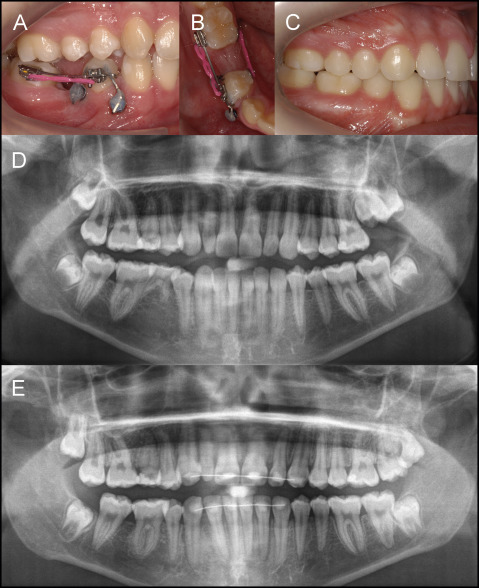
Skeletal anchorage for unilateral space closure was achieved by either miniscrews in the mandible (Aarhus Anchorage Screw System; American Orthodontics, Sheboygan, Wis) or palatal implants (SLA 4.2 mm long and 4.1 mm or 4.8 mm wide; Straumann, Basel, Switzerland). Palatal implants were placed in patients with agenesis in both jaws, whereas miniscrews were used in patients with agenesis only in the mandible. Thus, the posterior teeth of the agenesis side were mesialized over a space previously occupied by a deciduous second molar, which was expected to be approximately 9.83 ± 0.50 mm ( Fig ).

All patients had full fixed orthodontic appliances in both arches from second molar to second molar from the start of active treatment or after space closure. Thus, the molars were mesialized using either unilateral segmental fixed appliances in the mandible from first premolar to first molar or full fixed appliances in both arches from second molar to second molar (Roth prescription, slot size 0.022 in; American Orthodontics, Sheboygan, Wis) depending on the clinical condition. After initial tooth alignment, space closure was performed on 0.017 × 0.025-in or 0.019 × 0.025-in nickel-titanium wires. In patients with miniscrews, the mesialization force was exerted by elastomeric power chains, changed by the patients once every 2 weeks to prevent force decay. In patients with palatal implants, Class II intermaxillary elastics were used (1/4 in, 4.5 oz, changed every 12 hours). The regular recall appointment was every 6 to 8 weeks. Miniscrews were explanted after completion of space closure, whereas palatal implants were kept in place until the end of treatment.
For EARR measurement, all panoramic radiographs were scanned (Perfection V700 scanner; Epson America, Long Beach, Calif) with a resolution of 600 dpi at a scale of 1:1 and saved in portable network graphics format. Root length measurements were to be performed on the mandibular permanent canines, first premolars, and mesial and distal roots of the first molars on both sides of the mandible of each patient. For blinding reasons, each tooth was cropped from the panoramic radiograph (Paint Software; Microsoft, Redmond, Wash) and parameterized with random numbers between 0 and 1000 generated on https://www.random.org/ . The subsequent 300 tooth images were converted from portable network graphics format to tagged image file format (light image resizer 4 software, version 4.7.3.1; Pixmeo, Bernex, Switzerland) and imported into OsiriX Lite (version 6.5.2; http://www.osirix-viewer.com ), which was installed on an iMac 27-in computer (Mac OsX 10.6.8; Apple, Cupertino, Calif). The numbers of each radiograph linked with the patient data were saved in a separate file.
The first two authors performed all measurements independently within a week in a blinded manner, as described below. The tooth images were analyzed on the screen one by one. For better visualization of the tooth structures, the raters were allowed to adjust contrast and intensity and to enlarge the images as needed. First, the cementoenamel junction (CEJ) line was drawn by connecting the most mesial and distal CEJ points of each tooth. Then, the lines connecting the middle of the root apex with the corresponding cusp tip were drawn. In the permanent first molars, the distal root apex was connected with the distobuccal cusp and the mesial root apex with the mesiobuccal cusp. The distances between the cusp and apex and between the cusp and the CEJ line were measured on these lines with the “line caliper” function in the 2-dimensional viewer window of OsiriX and exported to an Excel (Microsoft) file. To overcome the difficulty of CEJ identification in certain molars, the distance from the most occlusal point of the molar furcation to the connecting line of the distal and buccal cusp tips was measured. This line was parallel to the dichotomous line of the mesial and distal roots long axes. This distance was used as an alternative crown length definition. The mean EARR measurement between the mesial and distal roots of each molar was used to define EARR in the molars.
All distances were measured in pixels. To convert pixels into millimeters, a 10-cm bar was scanned with the same units and settings used for the radiographs, and the length of the bar was measured in OsiriX in the same manner. Then, we used the subsequent equation 100 mm = 2409 pixels to convert pixels to millimeters in Excel ( Appendix ).
To control for distortion between the pretreatment and posttreatment radiographs caused by positional and angular changes of corresponding teeth due to treatment, a correction factor was used. Thus, the amount of EARR was calculated by the difference between the pretreatment and posttreatment radiographic lengths of the tooth multiplied by a correction factor. To determine this factor, crown length defined as the distance between the CEJ (intersection of the CEJ line and the line connecting the cusp with the apex), and the corresponding cusp tip (or the middle point between the distobuccal and mesiobuccal cusp tips for molars) was used as a constant and unchanging distance on the radiographs. Alternatively, crown length in molars was also defined using molar furcation, as described above, and the results were compared.
EARR was calculated using the following formula.
EARR = R 1 − [ R 2 × ( C 1 C 2 ) ]
Stay updated, free dental videos. Join our Telegram channel

VIDEdental - Online dental courses


