(1)
Canberra, ACT, Australia
Summary
The majority of endodontic failures can often be attributed to the inability of localising and treating all the canals of the root canal system. Root canal systems are commonly complex with the teeth often having lateral ramifications, extra roots or additional canals. Molar and premolar teeth can present with the highest incidence of aberrant morphology. A thorough knowledge of expected anatomy and variations from the norm are essential when undertaking root canal therapy to ensure success.
Clinical Relevance
The clinical impact of missed anatomy may result in failure and the necessity to carry out costly root canal re-treatment. Prevention of missed anatomy begins with a thorough knowledge of common tooth morphology. The clinician must be aware of the complexities of the root canal system and anatomical variations of the norm that may be encountered according to tooth type. The human dental pulp manifests multiple configurations and shapes which can vary from one individual to another. Careful interpretation of preoperative angled radiographs, correct access extension and a detailed exploration of the interior of the tooth using magnification and illumination are key steps for achieving clinical and radiographic success.
13.1 Overview of Root Canal Anatomy
In 1925 Hess and Zurcher first published a study showing us how the teeth had complicated root canal systems rather than the simplified canals that had been previously described [1]. For the first time we were able to see the intricate complexities of the internal root canal anatomy using vulcanised Indian rubber following dissolution of the surrounding tooth using 50 % hydrochloric acid. Even roots with a single canal will have lateral (an accessory canal located in the coronal or middle third of the root, usually extending horizontally from the main canal) and accessory canals leaving the main canal and communicating with the external surface of the root [2]. A thorough understanding of the complexities of the root canal system is essential for understanding the principles and problems of shaping and cleaning and for performing successful microsurgical procedures [3].
In order to grasp the varying anatomy that can occur with the teeth, a number of classification systems have been proposed. Weine’s classification of root canal morphology was based on the number of canal orifices, the number of canals and the number of foramina in each tooth [4] (see Fig. 13.1). A more comprehensive classification was provided by Vertucci, resulting in eight categories based on 2,400 extracted teeth that were rendered transparent and treated with a dye injection technique [5] (see Fig. 13.2). These classification systems provide us with a further insight into the complexities associated within the root canal system and help visualise the internal anatomy that we may become accustomed to encountering when carrying out preparation procedures.
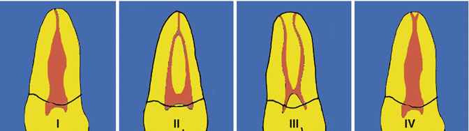
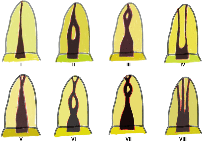
Fig. 13.1
Schematic diagrams representing Weine’s classification of root canal configurations. Type I – single canal from pulp chamber to apex. Type II – two canals leaving the chamber and merging to form a single canal short of the apex. Type III – two separate and distinct canals from chamber to apex. Type IV – one canal leaving the chamber and dividing into two separate and distinct canals
Fig. 13.2
Vertucci classification. Type I – a single canal with one foramen. Type II – two canals that join in the apical third. Type III – one canal that divides into two that subsequently reunites and exits as one. Type IV – two separate canals all the way to the apex. Type V – one canal that divides just short of the apex. Type VI – two canals that unite in the root and divide again at the apex. Type VII – one canal that divides, reunites and finally exits through two apical foramina. Type VIII – three separate canals in one root
Maxillary central and lateral incisors generally have one canal [3, 5]. When more than one canal is present, the possible configurations include two canals joining into one apical foramen (Vertucci type II) [6], two separate canals in one root (Vertucci type IV) [5], two canals in two separated roots or two or more canals associated with developmental abnormalities such as germination, fusion and dens invaginatus [7, 8]. Maxillary incisors with more than one root canal are rare and adequate access opening and observations of intra-operative radiographs should help in these difficult cases.
The majority of maxillary canine teeth have one canal in one root [5]. Morphological variations do exist but are rare where more than one canal is present [9].
The most common canal configuration for the maxillary first premolar is two separate canals in one root (Vertucci type IV) with a frequency of about 60–65 %. One canal that extends from the pulp chamber and dividing in the mid-root into two canals (Vertucci type V) can occur in 6–7 % of cases. In about 8–9 % of cases, the maxillary first premolar can have one canal and in 16–18 % two canals joining into one. On rare occasions three canals (mesio-buccal, disto-buccal and palatal) can occur in 2–6 % with a root disposition similar to the first permanent maxillary molar tooth [5, 10–12].
Maxillary second premolars usually have one canal in one root (Vertucci type I) in 38–48 % of cases, two canals joining in one root (Vertucci type II) in 20–22 % of cases and one canal separating into two canals that rejoin in the apical third in 5–10 % of cases (Vertucci type III). Two canals in two roots can occur in the region of 10–20 % of cases and one canal that splits and exits as two (Vertucci type V) in 6–9 % of cases. Rarely two canals that rejoin and split again (Vertucci type VI) occur in 2–5 % of cases. Three separate canals occur in the frequency of 1–2 %. Due to the aberrant canal morphology in these teeth, clinicians should pay close attention to both pre- and intra-operative radiographic examinations and clinical pulpal floor anatomy [5, 10–12].
A wide range of variation exists for the maxillary first permanent molar in the literature. It is generally accepted that this tooth has three roots (95 %) and four canals. Four percent of teeth have only two roots. The broad bucco-lingual dimension of the mesio-buccal root and associated concavities on the mesial and distal surface is consistent with the majority of mesio-buccal roots having two canals. The incidence of root fusion of two or three roots is approximately 5.2 %. Conical and C-shaped root morphology is very rare (<1 %). The incidence of two canals in the mesio-buccal root is 60 % and one canal to be about 40 %. The incidence of more than one disto-buccal or palatal root is rare (<1 %) [13–21].
The majority of maxillary second permanent molars have three roots with four root canals (90 %). Six percent may present with two roots and 3 % with a single root. Vertucci described the mesio-buccal root of having one canal in 71 % of cases (type I), two canals joining in 17 % (type II) of cases and two separate canals in 12 % of cases (type IV). The incidence of C-shaped canals is very rare in this subgroup of teeth [5, 15, 22].
The morphology of mandibular central and lateral incisors is not very dissimilar with the majority of teeth having two canals (25–40 %) [5, 23, 24].
The mandibular permanent canine tooth often has a single root (98 %) with a single canal and one apical foramen (92 %). Five percent of cases have two canals, which are confluent in the apical third forming one canal. The incidence of two canals and two separate roots is rare (<3 %) [5, 25].
A literature review of the root canal morphology of the mandibular first permanent premolar revealed that these teeth were one (98 %), two (1.8 %) and rarely three or four rooted (<0.1 %). Internal canal morphology revealed the majority of canals were single (76 % type I). Studies and case reports have also confirmed variation of canal morphology where division of the canal occurs in the middle or apical third into buccal and lingual branches (type II and IV). Rarely three canals can be present and occasionally the presence of C-shaped canals [5, 26–30].
The majority of mandibular second permanent premolars have a single root canal system (97.5 % type I) and two canals in the remainder (2.5 % type IV). The frequency of three of more canals is very rare (<0.4 % type VIII) [5, 29–31].
The majority of mandibular first permanent molars are often found to be two rooted with various canal configurations existing. Often two separate canals occur in 59 % of teeth (type IV), two canals conjoining apically in 28 % of teeth (type II) and a single canal system in 12 % of teeth. Occasionally three canals may be present in 1 % of teeth (type VIII). The frequency of the middle mesial canal varies between 1 and 7 % with variations of either conjoining in the middle or apical third or rarely with three separate exits apically. Distal roots often have one single canal in 70 % of teeth (type I), two canals that join into one in 15 % of teeth (type II), two separate canals in 5 % of teeth (type IV), one canal that divides into two in 8 % of teeth and one canal that divides into two then joins again in the apical third, forming a single canal in 2 % of teeth (type III). Overall the distal canal often presents with two or more canals in 30 % of cases. A major variant of root morphology known as radix entomolaris can occur rarely in 5 % of Caucasians but more often in populations with mongoloid traits (5–30 %) [5, 32–37].
Mandibular second permanent molars usually have two roots (mesial and distal). In the mesial root a type I canal configuration may be present (27 %), two canals which co-join apically (38 % type II) and two separate canals (38 % type IV). In the distal root, variations of type I (92 %), type II (3 %) and type IV (5 %) can be found. The incidence of a C-shaped canal system in this particular tooth group varies between 2 and 30 % and found often with a higher prevalence in the Chinese population [5, 38–42].
The root morphology and canal anatomy of maxillary and mandibular third molars is highly variable with often fusion involving one or more of the roots [43].
To ensure that all internal anatomy is correctly identified and treated, good preoperative radiographs, correct access cavity preparation, enhanced lighting, use of the dental operating microscope and controlled use of ultrasonic instruments are necessary tools that must be adopted. The clinical impact of missed anatomy is likely a failure in the future, resulting in either costly re-treatment or worse still extraction of the tooth. On the other hand, localisation and treatment of all canal anatomy will typically lead to a favourable outcome with complete clinical and radiographic healing. Cone-beam technology has been present for the last 30 years, but recent convergence and innovations have lead to the viable application of cone-beam volumetric tomography (CBVT) or cone-beam computed tomography (CBCT) in the dental office. Together these advantages have allowed CBVT to become a valuable tool in the modern endodontic practice, allowing us to truly appreciate the difficulties of treating these three-dimensional spaces that are more complex than we could have imagined [44–46] (see Chap. 12).
13.2 Maxillary Central Incisor Teeth
The root of the maxillary central incisor is often bulkier than the lateral with cross-sectional anatomy varying from triangular, circular or oval in shape. The canal is often tapering towards the apex in mature teeth with minimal curvatures in the apical portion (Fig. 13.3).
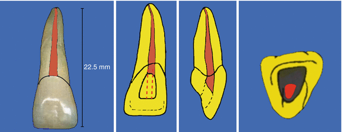
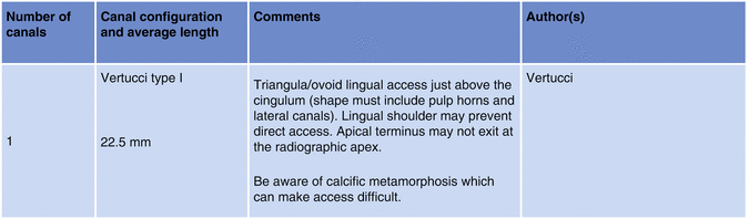
Fig. 13.3
Overview of maxillary permanent central incisor teeth
The access cavity is initiated by penetrating the bur occlusal to the cingulum, avoiding the incisal edge. Once penetration to the root canal is achieved, the access cavity must be refined in a mesio-distal direction to remove the entire roof associated with the pulp horns. The access cavity achieves a roughly triangular shape with this preparation, which mirrors the anatomy of the pulp chamber (see Fig. 13.4).
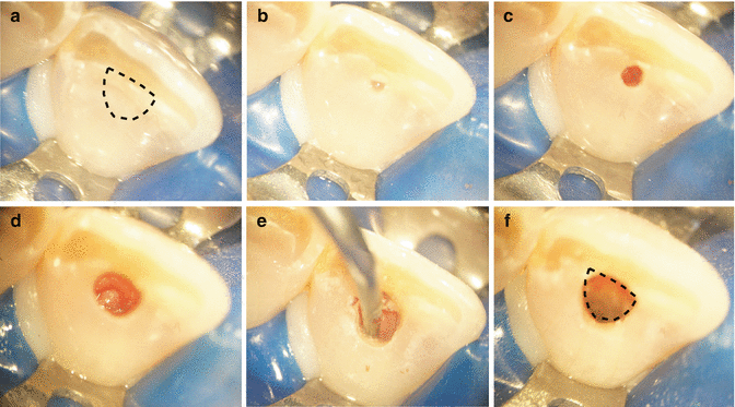
Fig. 13.4
Clinical photographs showing step-by-step access preparation in an anterior maxillary central incisor. Note (a) preoperative view (dotted line representing intended triangular access incorporating pulp horns), (b) initial bur penetration, (c) root canal penetrated, (d) widening of access in a mesial and distal direction to ensure pulp horns are incorporated, (e) use of ultrasonics to remove overhanging dentine and to ensure pulp horns are free of any tissue remnants and (f) final access preparation completed
Lateral canals may be present in the middle or apical third and the occurrence of a second canal is very rare. Due attention must be given to canals with very wide-open apices (blunderbuss teeth) and also narrow calcified canals (calcific metamorphosis) typically encountered following a traumatic incident. A sharp bend or curvature in the root would indicate previous trauma or bony interference during root formation (dilacerations), which may also affect management.
13.3 Maxillary Lateral Incisor Teeth
The lateral incisor is often smaller in comparison to its neighbouring central counterpart. The root is often slender with an apical curvature generally in a disto-palatal direction.
The presence of extra roots or occurrence of developmental grooves is more likely in this tooth group. Tooth invaginations (dens in dente) are also a common finding, which complicates endodontic management. Early recognition is essential and referral is probably best considered (Fig. 13.5).
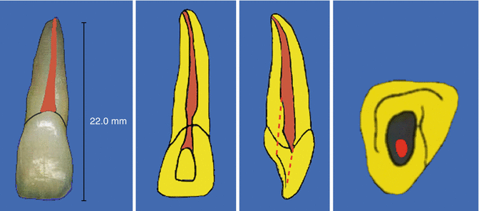
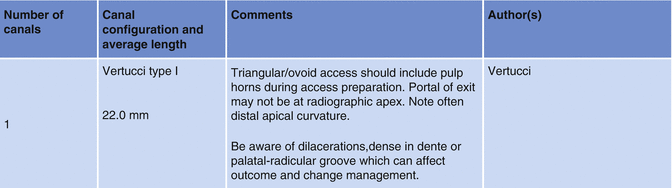
Fig. 13.5
Overview of maxillary permanent lateral incisor teeth
In this tooth, the access cavity is created in the same way as in the central incisor. The pulp horns are often closely situated or singular in this tooth, resulting in a final shape that is more likely to be ovoid as opposed to triangular. Care must be taken when negotiating the apical curvature in this tooth, particularly with larger file sizes, which can result in canal transportation or ledging if not correctly identified (see Fig. 13.6).
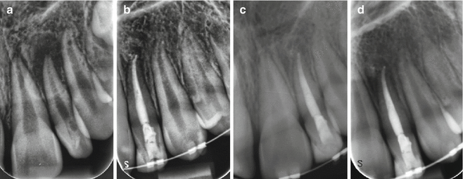
Fig. 13.6
Clinical radiographs demonstrating management of maxillary lateral incisor. Note the apical curvature that cannot be appreciated due to the distal and palatal curvature. (a) Preoperative view with large peri-radicular radiolucent lesion and (b) completed endodontic treatment of tooth 22. Note distal curvature of apical third maintained. Significant reduction of peri-radicular lesion achieved following chemo-mechanical debridement and interim intra-canal dressing of calcium hydroxide over a 3–month period, (c) 6–month follow-up and (d) 18–month follow-up showing continued healing
13.4 Maxillary Canine Teeth
These teeth can be one of the longest encountered with roots that are irregularly tapered and wide in a labio-palatal direction. The canal is typically a type I configuration with a root curvature apically often in a distal direction although can occur in any direction (Fig. 13.7).
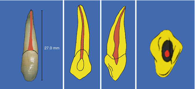
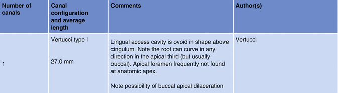
Fig. 13.7
Overview of maxillary permanent canine teeth
The access cavity begins about halfway up the crown on the palatal side (see Fig. 13.8). With an ovoid pulp chamber and a single horn, the access cavity is oval in outline and shape. The root canal is quite straight in the coronal and middle third and long enough to require the use of long 30-mm instruments.
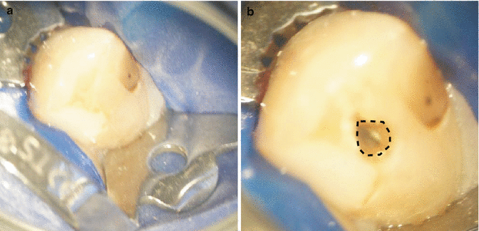
Fig. 13.8
Clinical photographs demonstrating access cavity preparation in a maxillary canine tooth. Note (a) preoperative view and (b) completed access preparation showing ovoid outline
Canines with a normal crown and root but with two canals are rare and difficult to identify because the canals may be superimposed. An adequate pulp chamber opening and careful observation of intra-operative radiographs may help in these difficult cases.
13.5 Maxillary Premolar Teeth
The maxillary first premolar has variable morphology but is generally considered to have two roots and two canals (see Fig. 13.9). The second premolar although variable at times generally presents with a single-rooted single canal system (see Fig. 13.10).
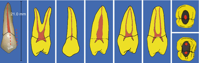
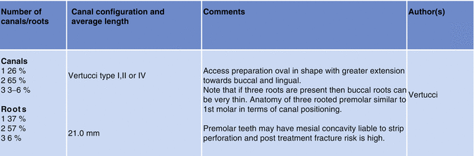
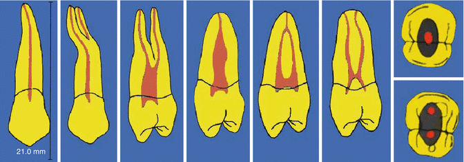
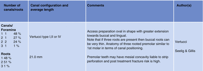
Fig. 13.9
Overview of maxillary permanent first premolar teeth
Fig. 13.10
Overview of maxillary permanent second premolar teeth
Careful radiographic assessment is essential in order to ascertain the likely number of roots and canal morphology before any operative intervention. Peri-apical radiographs give a two-dimensional image of a three-dimensional root canal system, so a minimum of two radiographs should be taken to reveal external and internal features of the tooth. A parallel radiograph should be taken with either a mesial or distal horizontal tube shift. The second angled radiograph may help visualise superimposed roots, displace the zygomatic process of the maxillary bone and increase the chances of observing additional root canals present.
The intra-oral radiograph should be carefully inspected for any sudden changes in the radiographic density of the root canal space, which may indicate bifurcation or trifurcation of the roots. Any sudden narrowing or disappearance may indicate branching of the root canal system often encountered in these teeth. The same principles can be applied during peri-operative treatment whereby any asymmetry of the file placed in the canal can also indicate the presence of a second root system.
The pulp chamber of the upper first and second premolar is oriented more towards the bucco-lingual direction since in the great majority of cases, it has two canals beneath the respective cusps (see Figs. 13.11 and 13.12). The point of entry of the bur is the middle of the central sulcus drilling parallel to the long axis of the tooth.
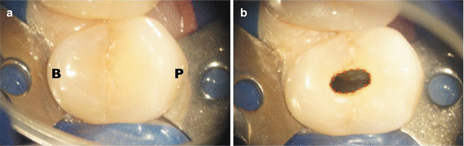
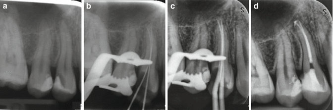
Fig. 13.11
Preoperative clinical photograph showing (a) occlusal anatomy of an upper maxillary premolar tooth and (b) completed ovoid access preparation extended in a bucco-palatal direction
Fig. 13.12
Clinical radiographs and photographs demonstrating endodontic treatment of a maxillary first premolar tooth with two canals. Note (a) preoperative radiograph demonstrating a two-rooted tooth, (b) MAF, (c) cone fit and (d) final obturation showing Vertucci IV canal configuration
The floor of the pulp chamber should be carefully examined looking at the position and symmetry of the canals. Asymmetry is often a good indication of additional anatomy that may be present. Upper premolars with three canals require a modified access cavity with a mesio-distal extension in the buccal portion of the traditional cavity. This modification, resulting in a T-shaped access, permits good access to both buccal canals. The presence of three canals is higher in the first premolar compared to the second but nevertheless rare. The anatomical roots are similar to the first molar whereby an MB, a DB and a P root may be present.
Treatment of maxillary premolars requires extra attention due to their extreme variability that can be encountered. Caution is advised due to the higher risk of failing to treat all internal anatomy, and a dental operating microscope or other forms of magnification are a necessary tool when dealing with these teeth.
13.6 Maxillary Molar Teeth
The literature shows a wide variation in anatomy of the maxillary molars although the average tooth often has three roots and four canals (see Figs. 13.13 and 13.14). The palatal root is often the longest with a curvature in the apical third towards the buccal. The disto-buccal root, which is shorter in comparison, may curve towards the mesial or distal in the apical third. The mesio-buccal root shows the greatest variation with a root that is broad in the bucco-palatal plane and narrowing in the mesio-distal place. This ribbon-shaped root will generally exit the crown mesially but can commonly abruptly curve towards the distal. Curvatures are often encountered in the apical third with possibilities of convergence and joining of additional mesio-palatal canals if present (see Fig. 13.15).
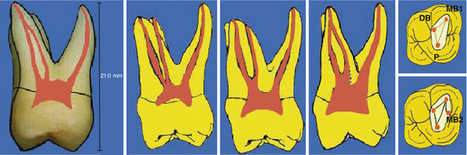
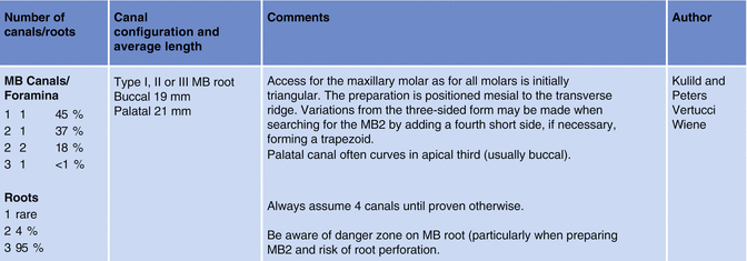
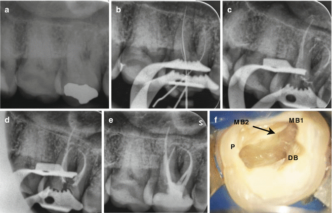
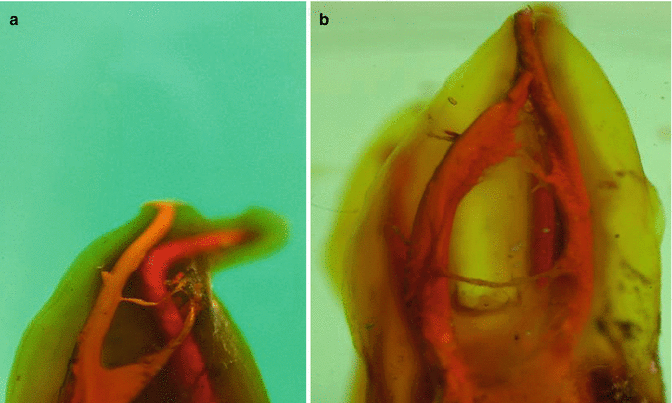
Fig. 13.13
Overview of maxillary permanent first molar teeth
Fig. 13.14
Clinical radiographs demonstrating root canal treatment carried out in a permanent maxillary first molar with three roots and four canals. Note (a) preoperative view, (b) IAF, (c) mid-fill and (d and e) final obturation using warm vertical compaction. (f) MB2 canal was located following ultrasonic troughing in this area (arrowhead)
Fig. 13.15
Clinical photographs demonstrating cleared maxillary first permanent molar teeth showing complex canal anatomy. (a) Maxillary first permanent molar showing the MB root. Note Vertucci classification VI (2–1–2) with a lateral canal from the MB1 canal. (b) Maxillary first permanent molar with two palatal canals. Note Vertucci classification II (2–1) with interconnecting fins and communication between both canals
Access preparation will be mesial to the external oblique ridge, allowing it to be maintained during preparation whereby enhancing the structural strength of the tooth. The orifice of the palatal canal is often the most prominent beneath the mesio-palatal cusp. The mesio-buccal canal often lies directly beneath the mesio-buccal cusp tip and the disto-buccal canal can often be found 2–3 mm distal to this canal and slightly towards the palatal. Careful inspection of the floor of the pulp chamber may reveal additional anatomy.
Recommended clinical approaches when treating maxillary molars include:
1.
Take two diagnostic radiographs, one parallel and one either with a mesial or distal tube horizontal tube shift, to correctly assess and identify external and internal anatomy.
2.
Careful removal of the pulp chamber roof is carried out using a non-end cutting to avoid damage to the pulpal floor. Dark developmental lines on the floor of the pulp chamber should be observed like a ‘road map’ to identify possible root canals present.
3.
Careful observation of the pulp chamber floor may also indicate additional root canal anatomy using sodium hypochlorite which may effervesce.
4.
Use of the dental operating microscope or dental loupes with adequate illumination is essential to correctly identify internal anatomy and avoid iatrogenic damage to the floor of the pulp chamber.
5.
Access cavity preparation does not need to be extended beyond the marginal ridge (see Fig. 13.16). Loss of marginal ridge greatly reduces the strength of the tooth.
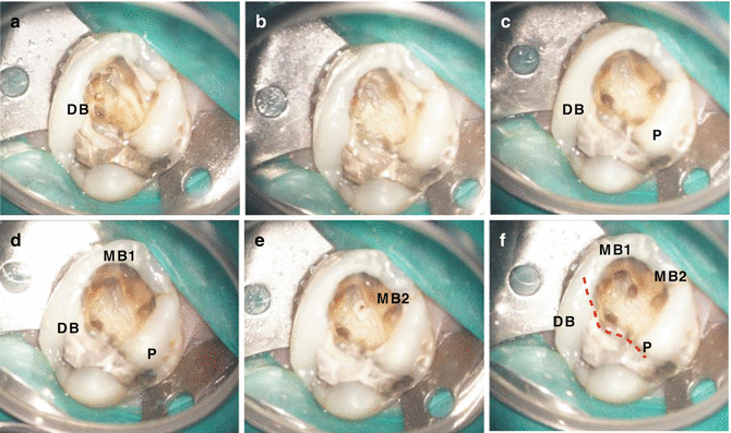
Fig. 13.16
Clinical photographs demonstrating access cavity preparation and refinement during the treatment of a maxillary permanent first molar. Note (a) calcified floor of pulp chamber with only DB canal located, (b) ultrasonic troughing carried out, (c) DB and P canal orifices located, (d) MB1 canal orifice located, (e) MB2 canal orifice located and (f) final four canals cleaned and shaped. Access cavity is not extended beyond marginal ridge (red dash line)
6.
Refinement of the access cavity will be required in the mesio-palatal direction using a rhomboid access. The MB2 canal is often palatal and often mesial in a line drawn between the MB1 and the palatal canal (see Fig. 13.16).
7.
Use of methylene blue dye stains can be useful to highlight the pulp chamber anatomy.
8.
Trough and search with a low-speed bur or ultrasonic tip beginning from the MB1 orifice. Be careful not to exceed a depth of 2–3 mm.
Stay updated, free dental videos. Join our Telegram channel
VIDEdental - Online dental courses


