Introduction
The aim of this study was to compare the palatal ruga patterns in subjects with oligodontia and normal tooth numbers.
Methods
An observational investigation was conducted by using maxillary dental study casts to compare ruga numbers, lengths, and shapes in subjects with diagnosed oligodontia or normal tooth numbers.
Results
A total of 32 subjects comprised both the oligodontia (mean age, 14.0 years; SD, 5.0 years) and the control (mean age, 14.5 years; SD, 5.1 years) groups. The mean number of missing teeth in the oligodontia group was 8.7. The mean number of rugae in the whole sample was 7.36 (SD, 1.16), with no significant difference between the groups ( P = 0.264). For ruga pattern, no differences were found for right-sided rugae; however, on the left side, a significant difference existed in shape frequency associated with ruga 2. Specifically, a curved shape was seen more frequently in ruga 2 of the oligodontia group ( P = 0.01).
Conclusions
The identification of subtle differences in ruga patterns between subjects with oligodontia and normal tooth numbers suggests potentially shared pathways during the development of these oral structures. Further large-scale investigations are warranted.
Graphical abstract
Highlights
- •
Palatine rugae form a series of transverse ridges in the hard palate mucosa.
- •
Their formation is regulated by molecular signaling pathways common to odontogenesis.
- •
We examined the relationship between ruga pattern and oligodontia in a pilot study.
- •
A significant pattern difference was found in subjects with oligodontia.
- •
Ruga pattern may provide evidence of underlying genetic variation.
The palatine rugae (rugae palatinae) are a series of irregular transverse ridges in the keratinized mucosa of the anterior hard palate, situated bilaterally adjacent to the midline incisive papilla and palatine raphe. The precise function of these structures is not fully understood, but their rich innervation suggests a role in mediating oral sensations, although they are also likely to perform a more physical function, facilitating manipulation of the food bolus in the oral cavity during mastication. Many species have palatine rugae, with specific numbers and patterns seen in different classes. In humans, the number of rugae is variable, ranging from 4 to 6 on each side, with a variety of different shapes and orientations having been described. The individuality, consistency of shape, and positional stability associated with these structures has led to their use in forensic science as an aid in human identification postmortem (palatoscopy) and as a reference point for the superimposition of serial models in orthodontics.
As a series of regularly spaced structures, the palatine rugae also offer insight into the biologic mechanisms that regulate anatomic periodicity. In the developing mouse embryo, the rugae are formed sequentially (with the exception of ruga 1), with the most posterior ruga (number 8) forming first, and successive new rugae (numbers 2-7) appearing anteriorly at sites of growth in the secondary palate. It has been demonstrated that a classic activator-inhibitor system is required during the sequential addition of these structures, mediated through interactions between the fibroblast growth factor and hedgehog signaling pathways. Moreover, an obligatory role for Wnt signaling has also been demonstrated during ruga development, potentially acting upstream of sonic hedgehog. It is now established that the same fundamental molecular signaling pathways actively regulate many aspects of development, including the craniofacial region. Interestingly, a significant number of genes identified in the developing rugae are also expressed in the developing dentition, and a potential genetic link between rugae and tooth development has been established in association with variations in the interferon regulatory factor 6 ( IRF6 ) gene in families with sporadic hypodontia. Selective tooth agenesis is a relatively common condition in humans, characterized by failure of tooth development. Incisor-premolar hypodontia, which often affects only 1 or 2 teeth, is the most common form of selective tooth agenesis; however, more severe forms are also seen, including hypodontia (absence of up to 6 teeth, excluding third molars) and oligodontia (absence of more than 6 teeth, excluding third molars). In recent years, a number of gene mutations have been identified in association with different forms of selective tooth agenesis, particularly oligodontia and including MSX1 , PAX9 , WNT10A , and AXIN2 .
In this preliminary study, we investigated the relationship between gross ruga pattern and tooth agenesis in untreated subjects with oligodontia and normal tooth number.
Material and methods
Subjects with oligodontia were identified by using retrospective searching of a hypodontia clinic database held in the orthodontic department at King’s College Hospital Dental Institute in London, United Kingdom. Unaffected control subjects were identified from those attending for routine orthodontic treatment. All subjects were white, over 8 years of age, and unrelated, and had complete records. There was no history of cleft lip and palate, orofacial syndromes, pathology, trauma, or surgery to the maxillary region, and no significant facial asymmetry or jaw discrepancy in any subject. In addition, oral hygiene was good, with no evidence of gingival or palatal inflammation. Those in the oligodontia group had 6 or more permanent teeth (excluding third molars) developmentally absent; those in the control group had all permanent teeth (excluding third molars) present. The presence or developmental absence of teeth was diagnosed after the clinical history and examination, supplemented with panoramic radiography taken as part of the routine care pathway for these subjects. The study was undertaken with approval from the Department of Research and Development, King’s College Hospital NHS Foundation Trust (reference number KCH14-009).
The sample size calculation was based on a chi-square test for the association between palatine rugae shape and normal (control) or genetically abnormal (oligodontia) groups. Assuming a 5% level of significance and 80% power, a total sample of 64 (minimum of 32 affected subjects and 32 controls) was required with an effect size of 0.5. This sample size calculation was carried out by using G*Power software (version 3.1; Heinrich Heine University, Dusseldorf, Germany).
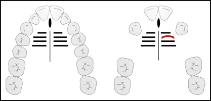
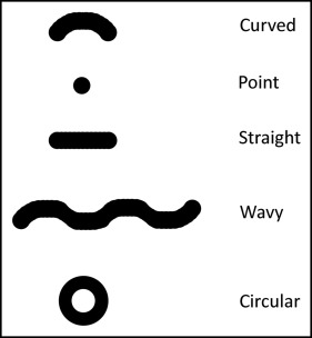
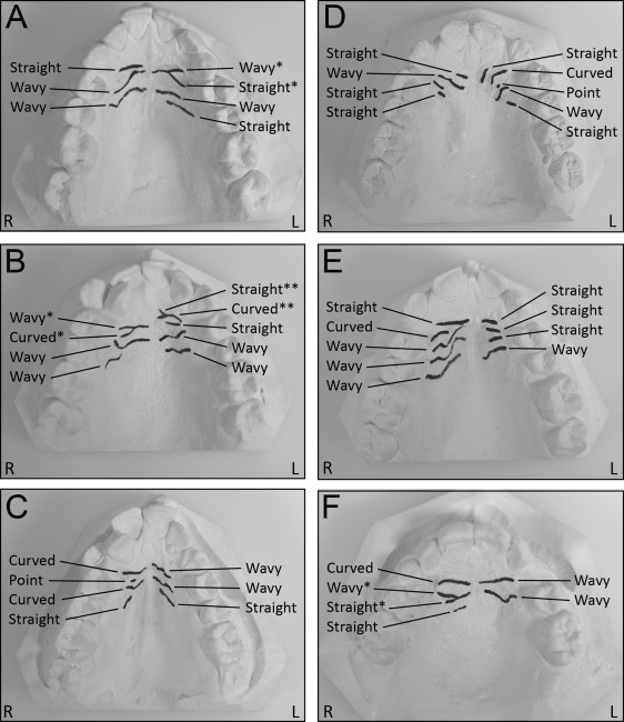
High-quality pretreatment dental stone (white orthodontic stone, ISO type 3; Whip Mix, Louisville, Ky) casts of the maxillary arch derived from alginate impressions and including the hard palate were available for all subjects. No orthodontic tooth movement had taken place in any subject. The palatal rugae were outlined with a sharp 6H pencil under suitable light, and magnification with the total number and sidedness (right or left) was recorded for each subject. The total length of each ruga from origin to termination (in millimeters) was measured using digital calipers accurate to within 0.01 mm, according to the manufacturer (ISO 9001, 150-mm electronic caliper; TESA Technology, Renens, Switzerland). For shape determination, the rugae were classified as curved, point, straight, wavy, or circular ( Fig 1 ). Curved types have a simple crescent shape that curves gently; point types are simple structures with no discernible shape and are less than 3 mm long; straight types run in a straight line from their origin to termination; wavy types are serpentine (if there was any curve at the origin or termination of a curved ruga, then it was classified as wavy); and circular types have a definite continuous ring. If there was any continuity between rugae (when individual rugae were connected either medially or laterally), the rugae were classified as divergent (having a single origin in the midline and diverging laterally) or convergent (having split origins and converging laterally); if there was no unification, rugae were classified as distinct ( Fig 2 ). All visible rugae were included in the analysis, irrespective of size. All rugae classifications and measurements were carried out by the same observer (A.M.). Ten records were reevaluated 1 week later to assess intraobserver reliability. A kappa coefficient of 0.62 demonstrated substantial agreement for measurements at the 2 time points; for shape classification, coefficients of 0.88 and 0.76 on the right and left sides, respectively, indicated good agreement.
Statistical analysis
Descriptive statistics are presented in means and standard deviations. The Mann-Whitney test was used to compare mean rugae values. Associations between rugae morphology (size and shape) and the presence or absence of oligodontia were tested by using the chi-squared test or the exact test. A P value of 0.05 was considered statistically significant.
Results
The total sample consisted of 64 subjects (48 female, 16 male) with a mean age of 14.3 years (SD, 5.0 years). The oligodontia group included 32 subjects (21 female, 11 male) with a mean age of 14.0 years (SD, 5.0 years), and the control group had 32 subjects (21 female, 11 male) with a mean age of 14.5 years (SD, 5.1 years). There was no significant difference in age distribution between the affected and unaffected subjects ( P = 0.54).
The mean number of missing tooth units in the oligodontia group was 8.7, and the distribution is shown in Figure 3 .
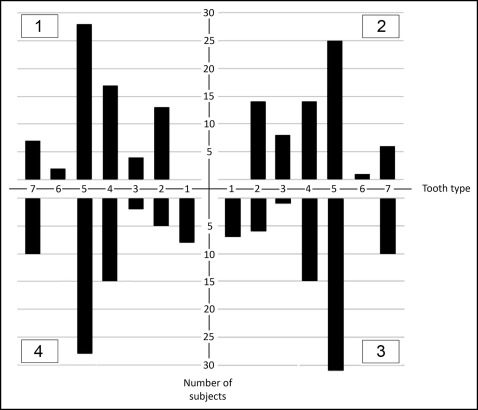
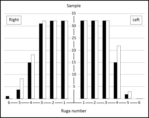
In the total sample, the mean number of rugae present was 7.36 (SD, 1.16). The distributions of the rugae are shown for both groups in Figure 4 . There was no significant difference in the mean numbers of rugae between the affected and unaffected subjects ( P = 0.10).
The mean lengths of the rugae are shown in Table I . There were no significant differences in mean rugae lengths between the samples. The distribution of ruga shapes (curved, point, straight, wavy, circular) are shown in Table II . There were no significant differences between the samples for right-sided rugae; however, on the left side, there was a statistically significant difference between the samples in shape frequency associated with ruga 2. Specifically, a curved shape was identified more frequently in ruga 2 of the oligodontia group ( P = 0.01). In addition, a wavy shape was more frequently associated with left ruga 3 in the control group, although this only approached significance ( P = 0.054). There were no significant differences in ruga unification between samples ( Table III ).
| Ruga number | Control group | Oligodontia group | P value | ||
|---|---|---|---|---|---|
| n | Mean length in mm (SD) | n | Mean length in mm (SD) | ||
| Right side | |||||
| 1 | 32 | 9.3 (2.6) | 32 | 9.0 (1.8) | 0.68 |
| 2 | 32 | 9.1 (3.8) | 32 | 9.9 (2.6) | 0.33 |
| 3 | 31 | 10.9 (3.7) | 32 | 10.7 (3.3) | 0.83 |
| 4 | 15 | 10.4 (3.7) | 18 | 9.2 (3.1) | 0.35 |
| 5 | 4 | 9.3 (5.3) | 8 | 9.3 (4.6) | 0.99 |
| 6 | 1 | 9.9 (-) | 0 | 0 (0) | – |
| Left side | |||||
| 1 | 32 | 10.0 (1.6) | 32 | 9.9 (1.5) | 0.76 |
| 2 | 32 | 9.8 (3.0) | 32 | 9.7 (2.4) | 0.85 |
| 3 | 32 | 11.2 (2.8) | 32 | 10.8 (3.0) | 0.60 |
| 4 | 15 | 10.1 (2.8) | 22 | 10.8 (2.6) | 0.44 |
| 5 | 2 | 11.3 (2.3) | 3 | 10.0 (1.6) | 0.52 |
| Ruga number | Group | Number of rugae | Curved | Point | Straight | Wavy | P value |
|---|---|---|---|---|---|---|---|
| Right side | |||||||
| 1 | Control | 32 | 15 | 0 | 10 | 7 | 0.17 |
| Oligodontia | 32 | 20 | 0 | 10 | 2 | ||
| 2 | Control | 32 | 8 | 2 | 8 | 14 | 0.34 |
| Oligodontia | 32 | 15 | 1 | 7 | 9 | ||
| 3 | Control | 31 | 6 | 1 | 4 | 20 | 0.90 |
| Oligodontia | 32 | 4 | 1 | 5 | 22 | ||
| 4 | Control | 15 | 2 | 0 | 4 | 9 | 0.12 |
| Oligodontia | 18 | 7 | 0 | 1 | 10 | ||
| 5 | Control | 4 | 2 | 0 | 1 | 1 | 0.50 |
| Oligodontia | 8 | 4 | 0 | 0 | 4 | ||
| 6 | Control | 1 | 0 | 0 | 0 | 1 | – |
| Oligodontia | 0 | 0 | 0 | 0 | 0 | ||
| Left side | |||||||
| 1 | Control | 32 | 21 | 0 | 3 | 8 | 0.14 |
| Oligodontia | 32 | 22 | 0 | 7 | 3 | ||
| 2 | Control | 32 | 9 | 0 | 8 | 15 | 0.01* |
| Oligodontia | 32 | 21 | 0 | 3 | 8 | ||
| 3 | Control | 32 | 5 | 0 | 3 | 24 | 0.054 |
| Oligodontia | 32 | 12 | 1 | 5 | 14 | ||
| 4 | Control | 15 | 1 | 0 | 3 | 11 | 0.27 |
| Oligodontia | 22 | 6 | 0 | 2 | 14 | ||
| 5 | Control | 2 | 1 | 0 | 0 | 1 | 1.0 |
| Oligodontia | 3 | 2 | 0 | 1 | 0 | ||


