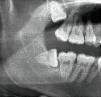
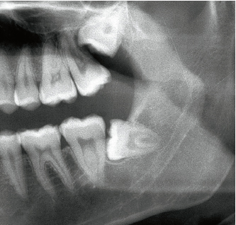
9-1b Ratio B crown-collar; Class II. The left mandibular third molar in the same patient.
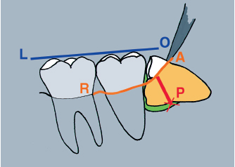
9-1c Ratio B. The crown of the left mandibular third molar is located below the occlusal line (OL). The impaction depth (P) is assessed in relation to the projection of the alveolar ridge (AR).
The impaction depth of the third molar is always assessed radiographically, starting from the alveolar ridge line (Figs 9-1a to 9-1c). The red line perpendicular to the bone contour in Fig 9-1c (P) indicates the real depth of impaction at the level of the mesial collar. Note that the occlusal line shows the orientation of the great axis of the second molar. The distal inclination of this tooth renders access more difficult, because in this case contact between the occlusal aspect and the distal wall of the second molar is very restricted.
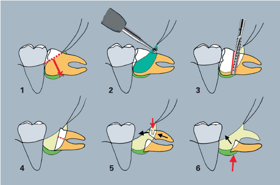
9-2 Surgical protocol for extraction of the left mandibular third molar, which has been retained horizontally. (1) The impaction depth is assessed starting from the mesial collar level. (2) The distal bone wall is easily accessed using a long round bur mounted on a handpiece. (3) The lateral removal of bone along the crown’s greatest contour and its sectioning are carried out using a spindle-shaped bur mounted on a contra-angle. (4) The root is separated using a long spindle-shaped bur mounted on a handpiece. The roots are then mobilized using a straight elevator. (5) A notch prepared with a fissure bur (red arrow) makes it easier to hold the distal root. (6) The mesial root is then tipped according to its curvature.
The surgical protocol for the extraction of a horizontal third molar remains more or less the same, whatever the depth of impaction (Fig 9-2). Three factors are taken into account: crown exposure, crown sectioning, and root extraction.
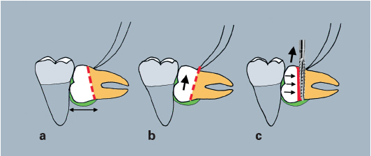
Crown exposure
The lining tissues are cut and reflected to obtain a large area of exposure of the buccal and lingual alveolar bone ridges in order to protect the soft tissues during crown sectioning.
Distal partial retention is reduced using a long perforating bur mounted on a handpiece. Drilling is facilitated when appropriate application points are used according to the axis of mouth opening. The exposure of the retromolar area extends up to the root into the spongy bone, 1 or 2 mm beyond the cementoenamel junction (see Fig 9-2, 2).
Lateral partial retention is exposed using the same round bur or a spindle-shaped bur mounted on a contra-angle. The greatest contour of the crown is exposed. It is not necessary to cut down the total height of the lateral wall of the crypt (see Fig 9-2, 2).
Stay updated, free dental videos. Join our Telegram channel
VIDEdental - Online dental courses


