Chapter 8
Review Visit: Problem Solving
Aim
This chapter discusses the more common problems associated with new complete replacement dentures and covers their diagnosis and management.
Outcome
By the end of this chapter, the practitioner should recognise that a range of problems may arise with new complete replacement dentures. The cause of these problems may relate to failure of the patient to adapt or to faults in the construction. If a well designed treatment plan has been followed by both the dentist and the dental technician, then many of these problems can be overcome with minor adjustments. Occasionally, a more significant error will become apparent at the first review visit, and this will require more radical alterations and maybe a remake. The practitioner should realise that problems such as pain and looseness may have a single underlying cause, and that eliciting the nature of the problem requires careful discussion with the patient and a thorough clinical examination.
The Review Visit
By the time of the first review visit, the patient should have worn the new dentures for approximately one week. The more common complaints reported include:
-
pain/discomfort
-
looseness of one or both dentures
-
speech problems
-
unsatisfactory appearance
-
chewing problems.
These problems are not necessarily mutually exclusive, for example pain can be associated with looseness of a denture. It is vital to listen carefully to the patient’s account as this is critical to accurate diagnosis and management of the problem. When the patient has outlined their concern, the clinician should conduct a clinical examination and attempt to relate the patient’s symptoms to the clinical signs.
Pain/discomfort
This is a particularly common finding with new dentures, and can have many causes.
Overextended periphery. Probably the most frequent aetiological factor is an area of overextension of the periphery. This should be suspected initially if there is an area of well-circumscribed soreness in the sulcus. This may appear as an area of erythema or ulceration of the mucosa. The clinician should relate the area of soreness to the periphery and subsequently adjust the offending area of overextension (as described in Chapter 7). This procedure is facilitated by using pressure-indicating paste (PIP). A small amount of PIP is applied to the suspected area of overextension and dabbed with a sponge. The denture is then reinserted, muscle trimmed and then removed. On inspection, the PIP will have rubbed off in the overextended area. This should be adjusted with a fluted acrylic trimming bur in a straight hand piece and the procedure repeated. When no more PIP rubs off, then the periphery should be polished. Usually, the patient will report instant improvement in discomfort. They should be advised that the soreness may take a day or so to disappear.
Freeway space problem. If there is pain across the entire lower denture-bearing area, then the patient may have too little freeway space. If the patient complains that this pain increases through the duration of the day, then it is almost certainly due to lack of freeway space. Speech may be affected, and further symptoms include unsatisfactory appearance and pain or tiredness in the jaw muscles. Freeway space can be increased by (a) raising the maxillary occlusal plane; (b) lowering the mandibular occlusal plane; or (c) a combination of both. The appearance of the dentures will help determine which of these options to choose. If, for example, there appears to be too much maxillary tooth showing, then option (a) should be chosen and the maxillary denture should be remade. In the case of the patient in Fig 8-1, both mandibular and maxillary dentures were at fault. The denture teeth should be removed from the denture base, a wax rim added and trimmed until sufficient freeway space is achieved. The jaw relationship in centric relation should be registered, and then a trial set-up prescribed in the usual manner. In some cases, there is too much freeway space and this may cause muscular discomfort. This problem will be described in more detail later.
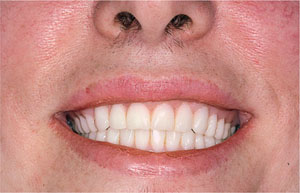
Fig 8-1 Patient with insufficient freeway space. Note, particularly, the level of the mandibular occlusal plane.
Pain in the sulcus. Sometimes, pain and ulceration in the sulcus cannot be attributed to the periphery of the denture. In this situation, the overextension may be in the lingual sulcus and is due to an error in the occlusion. The movement of the denture base, when the lower jaw moves from centric relation to the intercuspal position (ICP) should be noted. If maximal intercuspation is not evident in centric relation, then either a protrusive or lateral slide will occur. If a protrusive slide is present, this is likely to cause the denture base to move anteriorly and thus cause trauma to the anterior lingual sulcus. If a lateral slide is evident, then contact occurs on one side and the denture base will move to the contralateral side, i.e. a right-sided early contact will result in pain in the left lingual sulcus. If there is a large error (approximately half a tooth out), then the lower posterior teeth should be removed and the jaw relationship re-recorded. If the error is smaller, then clinical remount and adjustment on a semi-adjustable articulator should be undertaken, as described in Chapter 7.
Pain on crest of the alveolar ridge. This may be due to poor quality support tissues or the unemployed ridge phenomenon (described in Chapter 3). Prominent areas of bone have thin mucosa overlying them, and these offer poor support for the denture. They may respond well to relieving the corresponding areas of the denture. Marking the area with an indelible ink or drying the mucosa and applying PIP will help identify the corresponding area on the denture. When the area is sufficiently relieved, the clinician should be able to apply gentle digital pressure to the denture without causing pain. A further possible treatment option i/>
Stay updated, free dental videos. Join our Telegram channel
VIDEdental - Online dental courses


