Obtaining and Interpreting Radiographs
QUESTIONS
1. A periapical (PA) image is needed on the maxillary left premolar area. The patient presents with a large maxillary torus. Using the paralleling, or right-angle, technique, the image receptor film or sensor is to be placed
A distal to the maxillary premolar area.
B mesial to the maxillary premolar area.
2. A periapical image of the maxillary right molar area is needed, but patient has a shallow palate, and the first image taken misses the apices of the teeth. To correct this error, using the paralleling technique, an acceptable image can be obtained by moving the position indicating device (PID) in a
A 20-degree difference in the horizontal angulation toward the mesial surfaces.
B 20-degree difference in the horizontal angulation toward the distal surfaces.
C positive 20-degree difference in the vertical angulation.
D negative 20-degree difference in the vertical angulation.
E direction in which the central ray is perpendicular to the image receptor.
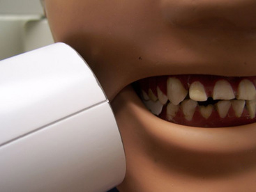
3. When taking a premolar bitewing image, which error in the position of the PID is seen in the photograph shown?

A Greater horizontal angulation than required
B Greater positive vertical angulation than required
4. The error that will occur to the premolar bitewing image as a result of the PID positioning will be
A unequal distribution of maxillary and mandibular teeth. There will be more coverage of maxillary teeth and fewer mandibular teeth.
B unequal distribution of maxillary and mandibular teeth. There will be more coverage of mandibular teeth and fewer maxillary teeth.
5. In the molar bitewing image, the error that will occur as a result of the PID positioning will be distal overlap. Distal overlap is corrected by repositioning the horizontal angulation of the PID more mesiodistally.
C The first statement is true, and the second statement is false.
D The first statement is false, and the second statement is true.
6. Considering exposure time during the production of x-rays, how many impulses are equivalent to 1⁄4 second?
7. Order the steps for the manual processing of x-ray films. Match each letter with its proper sequence number.
| Sequence Number | Steps |
| 1. _____ | A. Rinsing in water for 30 seconds |
| 2. _____ | B. Washing in water for 20 minutes |
| 3. _____ | C. Developing |
| 4. _____ | D. Fixing |
| 5. _____ | E. Drying |
8. From the following list, select the three elements used in the x-ray tube.
9. Which of the following is the unit used to measure radiation exposure?
10. A radiographic image will have increased density if the operator
C decreases the milliampere (mA).
D increases the distance of the x-ray source to the image receptor.
11. The smaller the silver halide crystals in the emulsion of an intraoral film, the faster the film speed. Faster film speed reduces radiation exposure to the patient.
C The first statement is true, and the second statement is false.
D The first statement is false, and the second statement is true.
12. Which ingredient of the developing solution in the manual processing of x-ray films is alkaline and aids in the softening of the emulsion?
13. In which anomaly is cementum of two adjacent teeth joined together?
14. Which structure or material will appear the MOST radiopaque on a dental image?
15. Which structure appears as a radiopaque line surrounding the root of the tooth?
16. Which of the following describes the appearance of a carious lesion on the buccal or lingual surface of a molar on a dental image?
17. Which mandibular anatomic landmark can be seen as a radiolucent area on a mandibular premolar periapical (PA) image?
18. All of the following are descriptions of a ghost image on a panoramic image EXCEPT one. Which one is the EXCEPTION?
A It is a similar sharpness as the original artifact.
B It is a similar shape to the original artifact.
C It is on the opposite side of the original artifact.
19. Cone cutting has occurred on the coronal portion of a mandibular anterior PA image. The operator will correct this error by
A moving the image receptor more superiorly.
B moving the image receptor more inferiorly.
C moving the PID to completely cover the image receptor.
20. The tubehead of the panoramic unit is angled so that the x-ray beam is directed
A in a slightly positive vertical angulation.
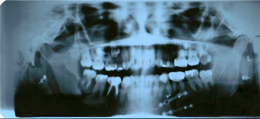
B in a strong positive vertical angulation.
C in a slightly negative vertical angulation.
21. From the following list, select the three statements associated with digital imaging.
A Sensors cannot be sterilized.
B A digital image is a two-dimensional representation of a three-dimensional object.
C Exposure times are 50% to 90% more than in traditional radiography.
D Digital imaging must be wired to function correctly.
E Assembled pictorial information with each gray value is assigned a digit in binary code.
22. The tubehead is a component of the x-ray machine that helps limit radiation to the patient in all of the following functions EXCEPT one. Which one is the EXCEPTION?
A Inherent filtration within glass tube, insulating oil, and tubehead
B Added aluminum disk filtration
C Production of both long-wavelength and short-wavelength x-rays
D Collimation with lead disk placed in the pathway of the x-ray beam
23. Erythema, nausea, diarrhea, hemorrhage, and loss of hair are signs and symptoms seen with
24. Which statement BEST describes changes to the atom during the ionizing process?
A The atom gains an electron and will have a negative charge.
B The atom gains an electron and will have a positive charge.
C The atom loses an electron and will have a negative charge.
D The atom loses an electron and will have a positive charge.
25. For each numbered anatomic landmark, select the BEST associated radiographic description.
| Anatomic Landmark | Description of Radiographic Presentation |
| ___1. Median palatine suture | A. Ring-shaped radiopacity noted apical to mandibular incisors |
| ___2. Genial tubercles | B. Dense radiopaque band extending from the molar region downward toward the premolar area of the mandible |
| ___3. Incisive foramen | C. Thin radiolucent line between maxillary central incisors |
| ___4. Mylohyoid ridge | D. Triangular-shaped radiopacity superimposed over or seen inferior to the maxillary tuberosity area |
| ___5. Mental foramen | E. Small oval or round radiolucency seen in the apical region of mandibular premolars |
| ___6. Floor of nasal cavity | F. Dense radiopaque band of bone seen apical to maxillary incisors |
| ___7. Coronoid process | G. Small oval or round radiolucency seen between the roots of maxillary central incisors |
26. Order each tissue in order of radiosensitivity, from most sensitive to least sensitive. Match each letter with its proper sequence number.
| 1. _____ | A. Skin |
| 2. _____ | B. Oral mucosal epithelium |
| 3. _____ | C. Nervous tissue |
| 4. _____ | D. Reproductive tissue |
| 5. _____ | E. Lymphatic tissue and bone marrow |
| 6. _____ | F. Mature bone and cartilage |
27. Which factor affects contrast?
28. For each numbered x-radiation interaction, select the BEST description.
| X-Radiation Interaction | Description |
| _____ 1. X-ray photon passed through atom unchanged; no interaction | A. Electrons stop suddenly as they hit the anode. |
| _____ 2. Coherent scatter | B. Ionization takes place when the photon ejects an outer shell electron. This results in the loss of energy for the photon and a negative charged electron. The low energy photon changes direction. |
| _____ 3. Photoelectric effect | C. An unstable atom releases energy. |
| _____ 4. Compton scatter | D. This is responsible for producing the densities on the receptor or film. |
| _____ 5. Bremsstrahlung radiation | E. Ionization takes place when photons are absorbed by patient tissues. |
| _____ 6. Radioactivity | F. No loss of energy and no ionization occur when this low-energy photon interacts with an outer shell electron. The unaffected photon changes direction. |
29. Order the steps in the production of x-radiation. Match each letter with its proper sequence number.
| 1. _____ | A. Electrons travel across to the anode when the high voltage circuit is activated. |
| 2. _____ | B. Thermionic emission occurs. |
| 3. _____ | C. 110 or 220 line voltage is reduced to 3 to 5 volts. |
| 4. _____ | D. Kinetic energy is converted to x-ray energy and heat. |
30. For each numbered term listed below, select the BEST description from the list provided.
| Term | Description |
| _____ 1. Anode | A. Produces electrons |
| _____ 2. Cathode | B. Controls the number of electrons |
| _____ 3. Kilovoltage | C. Decreases voltage |
| _____ 4. Milliamperage | D. Narrows the beam and controls the direction of the beam |
| _____ 5. Molybdenum cup | E. Release of electrons from the tungsten filament when the electrical current passes through it and heats the filament |
| _____ 6. Tungsten filament | F. Spot where the electrons are converted into x-ray photons |
| _____ 7. Tungsten target | G. Positive electrode |
| _____ 8. Thermionic emission | H. Process of producing electrons when a current is passed through tungsten wire |
| _____ 9. Step-down transformer | I. Controls the force of the electrons |
| ____ 10. Collimator | J. Negative electrode |
31. In the formation of x-radiation, electrons that strike the nucleus of a tungsten atom produce which type of x-ray?
32. You wish to compare your patient’s current images with images that were taken 2 years ago. The images from 2 years ago have a yellowish-brown color. Which of the following is the cause of the yellow-brown stains?
33. Which of the following is the receptor that captures computerized images as discrete units of information?
34. It is acknowledged that the routine performance of radiography is not recommended. Using sound professional judgment, for each numbered type of patient listed, select the recommended prescription of radiography based on the Guidelines for Prescribing Radiographs established by the U.S. Food and Drug Administration (FDA) from the list provided. Not all guidelines will be used.
| Type of Patient | Guideline for Prescribing Radiography |
| ____ 1. Recall patient: adult patient with no sign of clinical caries; presents with no caries risk | A. Posterior bitewings; every 6 to 18 months |
| ____ 2. Recall patient: child with no significant clinical caries; presents with no caries risk | B. Clinical judgment based on patient needs |
| ____ 3. New patient: 14-year-old with clinical caries in select areas | C. Every 3 to 6 months |
| ____ 4. Recall patient: adult with clinical caries or caries risk | D. Every 12 to 24 months if proximal contacts are not visible and cannot be checked with probe |
| ____ 5. Recall patient: 14-year-old with no sign of clinical caries; presents with no caries risk | E. Posterior bitewings with panoramic radiograph or posterior bitewing with select periapical radiography |
| ____ 6. Recall patient: adult patient with periodontal disease | F. Every 3 to 5 years |
| G. Every 18 to 36 months | |
| H. Posterior bitewings every 24 to 36 months | |
| I. Full mouth set of radiographs including bitewings |
35. Which of the following concepts are examples of the ALARA concept? (Select all that apply.)
A Compliance with maximum permissible dose of radiation for the occupationally exposed worker
B Use of evidence-based selection criteria for radiography for patient needs
36. From the following list, select the four characteristics that describe properties of dental x-radiation.
B May cause certain substances to fluoresce
C Travels at the speed of sound
37. Radiation may damage cells indirectly by damaging the cell nucleus. Direct cellular damage occurs when ionization causes radiolysis of water producing hydrogen peroxide.
C The first statement is true, and the second statement is false.
D The first statement is false, and the second statement is true.
38. When an x-ray photon passes through matter, which of the following situations is MOST likely to occur?
39. From the following list, select examples of stochastic effects of radiation exposure. (Select all that apply.)
40. Radiation exposure is not of concern when it occurs in small doses. The cumulative effect of radiation exposure may lead to health problems such as cancer.
C The first statement is true, and the second statement is false.
D The first statement is false, and the second statement is true.
41. All of the following are examples of radiosensitive cells EXCEPT one. Which one is the EXCEPTION?
42. Failing to follow radiation exposure patient guidelines would put a fetus at greatest risk in which trimester?
43. Which of the following BEST describes the process of heating the cathode wire until it is red hot and “boiling off” of electrons?
44. All of the following decrease radiation exposure to the patient EXCEPT one. Which one is the EXCEPTION?
B F-speed film in place of D-speed film
C 8-inch PID in place of 16-inch PID
45. Which of the following is (are) the maximum permissible dose (MPD) for a dental hygienist? (Select all that apply)
46. A radiograph that has many shades of grade is considered to have
47. The mental fossa is visible in which radiographic image?
48. All of the following phrases are consistent with the paralleling technique EXCEPT one. Which one is the EXCEPTION?
B It is the preferred method for taking radiographs.
C The film is placed parallel to the long axis of the tooth.
D The central ray is directed perpendicular to the tooth and the image receptor.
E It produces images with the least magnification and dimensional distortion.
49. Which radiolucent landmark is visible on a maxillary posterior periapical image?
50. To prevent overlapped contacts on periapical and bite-wing images,
A position the image receptor parallel to the long axis of the tooth.
B increase vertical angulation.
C decrease vertical angulation.
D direct the x-ray beam through the interproximal contacts.
E place the image receptor to include the correct teeth on the radiograph.
51. The radiographic appearance of the zygomatic process of the maxilla appears as a J-shaped or U-shaped radiopacity located superior to the maxillary first molar region. The visibility of the zygomatic process of the maxilla on the image is a result of utilization of the bisecting technique.
C The first statement is true, and the second statement is false.
D The first statement is false, and the second statement is true.
52. In the image shown, which permanent tooth is unerupted?
53. The coronoid process of the mandible appears as a triangular-shaped radiopacity. This marked prominence of bone found on the anterior ramus of the mandible is not seen on a mandibular periapical radiograph.
C The first statement is true, and the second statement is false.
D The first statement is false, and the second statement is true.
54. From the following list, select two structures that appear radiolucent on a radiographic image.
55. Which of the following techniques would be required to correct a nondiagnostic bitewing radiograph with overlapped interproximal contacts?
A Increasing the vertical angulation of the PID
B Decreasing the vertical angulation of the PID
56. Which of the following statements BEST describes the anomaly in the image shown below?
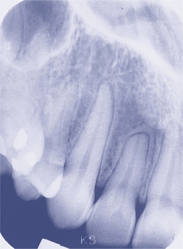
From Haring JI, Lind LJ: Radiographic interpretation for the dental hygienist, Philadelphia, PA, 1993, Saunders.
Stay updated, free dental videos. Join our Telegram channel
VIDEdental - Online dental courses


