Investigations
Informed consent and confidentiality are required for all investigations, and the advantages should clearly outweigh any dangers or disadvantages.
Blood tests
Blood tests can help determine disease states, but should be requested only when clinically indicated. There is always a danger of needlestick injury. Furthermore, abnormal ‘blood results’ do not always mean disease and false-positive results are possible. Serum is used for assaying antibodies, which can help diagnose infections and autoimmune disorders, and for assaying most biochemical substances (e.g. ‘liver enzymes’).
Microbiological tests
Microbiological diagnosis is based on either demonstration of the microorganism or its components (antigens or nucleic acids) directly in samples or tissues, which are best used as results are speedily obtained. The demonstration in the serum of a specific antibody response can be helpful.
Salivary flow determination
Unless the baseline salivary flow rate for an individual patient is known, it is impossible to be certain if there has been a reduction in salivary flow, since the salivary flow rate varies widely from person to person. Salivary flow rates also vary over time and so estimates should be taken on several occasions.
Normal and reference values are shown in Table 7.1.
Table 7.1
Whole saliva flow rates* (ml/min)
| Normal | Hyposalivation | |
| Unstimulated (resting) | 0.3–0.4 ml/min | <0.1 ml/min |
| Stimulated | 1–2 ml/min | <0.5 ml/min |
*Unstimulated salivary flow rate (USFR) measurement of whole saliva uses a simple draining test for 5 minutes at rest: a rate less or equal to 0.1 ml/min suggests hyposalivation.
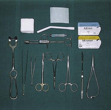
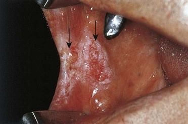
Biopsy
Before beginning, it is important to decide whether an excisional or incisional biopsy is indicated.
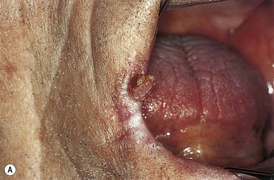
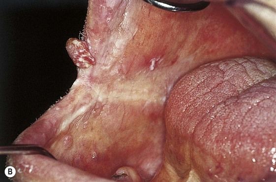
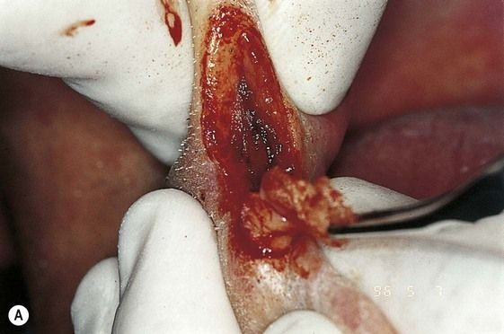
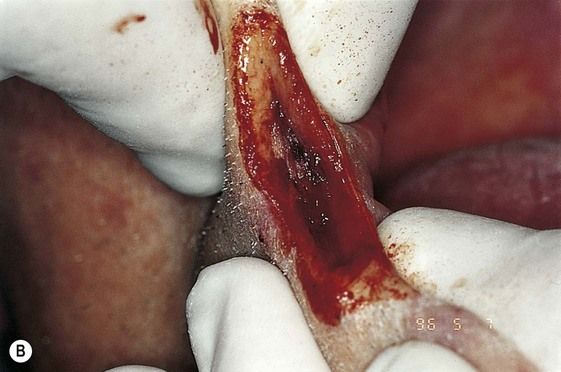
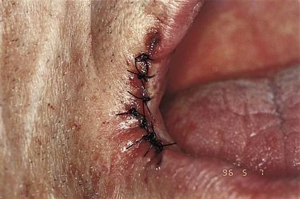
Excisional biopsy
Excisional biopsy (Figures 7.1–7.4) is used for small superficial lesions (max 1 cm). If a malignant tumour is suspected, even if small, an excisional biopsy must be done by a specialist.
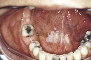
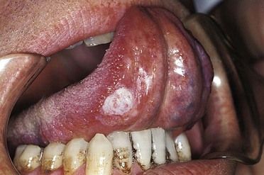
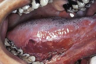
Incisional biopsy
Choose the area to biopsy based on:
An incisional biopsy (Figures 7.5–7.13) can be performed with a scalpel, or dermatology punch.

Stay updated, free dental videos. Join our Telegram channel
VIDEdental - Online dental courses


