General Physiology
Learning Outcomes
On completion of this chapter, the student will be able to achieve the following objectives:
• Pronounce, define, and spell the Key Terms.
• Name and locate each of the 10 body systems.
• Explain the purpose of each body system.
• Describe the components of each body system.
• Explain how each body system functions.
• Describe the signs and symptoms of common disorders related to each body system.
• Give examples of conditions that require interaction among body systems.
Electronic Resources
 Additional information related to content in Chapter 7 can be found on the companion Evolve Web site.
Key Terms
Apocrine sweat glands Large sweat glands that are found under the arms, around the nipples, and in the genital region.
Appendicular (ap-en-DIK-yoo-lur) skeleton Portion of the skeleton that consists of the upper extremities and shoulder girdle plus the lower extremities and pelvic girdle.
Arteries Large blood vessels that carry blood away from the heart.
Articulation (ahr-tik-yoo-LAY-shun) Another term for joint.
Axial (AK-see-ul) skeleton Portion of the skeleton that consists of the skull, spinal column, ribs, and sternum.
Bone marrow Gelatinous material that produces white blood cells, red blood cells, or platelets.
Cancellous (KAN-sil-us) bone Lightweight bone found in the interior of bones; also known as spongy bone.
Capillaries A system of microscopic vessels that connects the arterial and venous systems.
Cartilage Tough, connective, nonvascular elastic tissue.
Central nervous system (CNS) The brain and spinal cord.
Compact bone Outer layer of bones, where needed for strength; also known as cortical bone.
Gomphosis A type of fibrous joint such as a tooth into the alveolus.
Integumentary (in-teg-yoo-MEN-tuh-ree) system The skin system.
Involuntary muscles Muscles that function automatically without conscious control.
Joints Structural areas where two or more bones come together.
Muscle insertion Location where the muscle ends; the portion away from the body’s midline.
Muscle origin Location where the muscle begins; the portion toward the body’s midline.
Neurons Direct nerve impulses.
Osteoblasts (OS-tee-oe-blasts) Cells that form bone.
Pericardium (per-i-KAHR-dee-um) Double-walled sac that encloses the heart.
Periosteum (per-ee-OS-tee-um) Specialized connective tissue that covers all bones of the body.
Peripheral nervous system (PNS) Cranial nerves and spinal nerves.
Peristalsis (per-i-STAHL-sis) Rhythmic action that moves food through the digestive tract.
Plasma A straw-colored fluid that transports nutrients, hormones, and waste products.
Red blood cells Cells that contain the blood protein hemoglobin, which plays an essential role in oxygen transport; also known as erythrocytes.
Sebaceous glands Oil glands that keep the hair and skin soft and are associated with sex hormones.
Sharpey’s fibers Tissues that anchor the periosteum to the bone.
Sudoriferous (soo-doe-RIF-ur-us) glands Sweat glands that are widely distributed over the body and provide heat regulation.
Veins Blood vessels that carry blood to the heart.
White blood cells Cells that have the primary function of fighting disease in the body; also known as leukocytes.
The human body is the most incredible creation, with its senses and strengths, an ingenious defense system, and mental capabilities. The human body is a masterpiece more amazing than science fiction.
The study of the human body is as old as human history, because people have always had a fascination about how the body is put together, how it works, why it becomes diseased, and why it wears out.
Physiology is the study of how living organisms function. It continues beyond the study of anatomy into how the body works, what it can do, and why (see Chapter 6).
The human body has 10 systems: (1) skeletal, (2) muscular, (3) cardiovascular (including lymphatic and immune systems), (4) nervous, (5) respiratory, (6) digestive, (7) endocrine, (8) urinary, (9) integumentary (skin), and (10) reproductive. Each system has specific organs within it, and each body system performs specific functions. When all 10 systems are functioning well, the person is healthy (Table 7-1). This chapter summarizes each of these body systems, their components and functions, and the disorders that affect them.
TABLE 7-1
| Body System | Components | Major Functions |
| Skeletal system | 206 bones | Protection, support, and shape; hematopoietic; storage of certain minerals |
| Muscular system | Striated, smooth, and cardiac muscle | Holding body erect, locomotion, movement of body fluids, production of body heat, communication |
| Cardiovascular system | Heart, arteries, veins, blood | Respiratory, nutritive, excretory |
| Lymphatic and immune systems | White blood cells; lymph fluid, vessels, and nodes; spleen and tonsils | Defense against disease, conservation of plasma proteins and fluid, lipid absorption |
| Nervous system | Central and peripheral nervous systems, special sense organs | Reception of stimuli, transmission of messages, coordinating mechanism |
| Respiratory system | Nose, paranasal sinuses, pharynx, epiglottis, larynx, trachea, bronchi, and lungs | Transport of oxygen to cells, excretion of carbon dioxide and some water wastes |
| Digestive system | Mouth, pharynx, esophagus, stomach, intestines, and accessory organs | Digestion of food, absorption of nutrients, elimination of solid wastes |
| Urinary system | Kidneys, ureters, bladder, and urethra | Formation and elimination of urine, maintenance of homeostasis |
| Integumentary system | Skin, hair, nails, and sweat and sebaceous glands | Protection of body, regulation of body temperature |
| Endocrine system | Adrenals, gonads, pancreas, parathyroids, pineal, pituitary, thymus, and thyroid | Integration of bodily functions, control of growth, maintenance of homeostasis |
| Reproductive system | Male: testes, penisFemale: ovaries, fallopian tubes, uterus, vagina | Production of new lifeProduction of new life |
Skeletal System
The skull, spine, and rib cage form the axial skeleton and account for 80 of the 206 bones in the human body. The shoulders, arms, hands, hips, legs, and feet form the appendicular skeleton. The skull consists of 28 bones and is discussed in detail in Chapter 9 (Fig. 7-1).
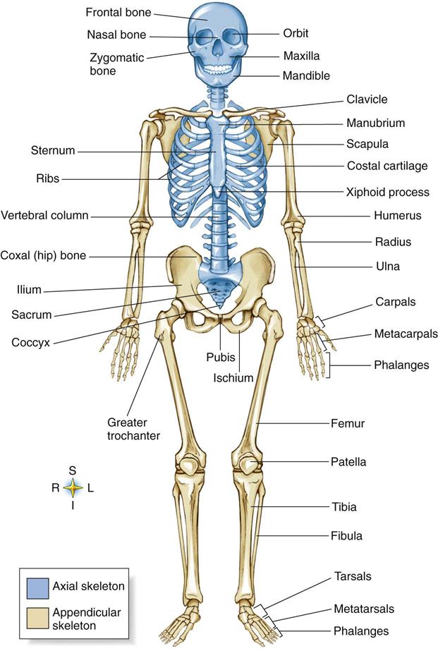
The axial skeleton (80 bones) consists of the skull, spinal column, ribs, and sternum. Its function is to protect the major organs of the nervous, respiratory, and circulatory systems.
The appendicular skeleton (126 bones) consists of the upper extremities and shoulder area plus the lower extremities and pelvic area. It protects the organs of digestion and reproduction.
Many disorders can affect the skeletal system (Table 7-2).
TABLE 7-2
Disorders of the Skeletal System
| Disorder | Description | Signs and Symptoms |
| Arthritis | Inflammation of a joint. More than 100 forms are known, and all have different causes. | Swelling and pain, usually with structural changes. Mobility impairment and difficulty in performing daily tasks |
| Fracture | Broken bones caused by stress on the bone. Fractures can occur in any bone in the body and are classified by type. | Severe pain, swelling, and disfigurement, depending on the type of fracture |
| Gout | Inflammatory joint reaction caused by accumulation of uric acid crystals. The area that is usually affected is the big toe. | The joint usually becomes red, warm, shiny, swollen, and very sensitive to the touch. |
| Osteomyelitis | Infection of the bone that is caused by bacteria, fungi, or contaminated foreign material such as an artificial joint | Sudden onset of fever, limited movement, and severe pain in the body part involved |
| Osteoporosis | Age-related disease in which bone is demineralized, resulting in loss of bone density and fracture | Frequent fractures, especially of the vertebrae, wrist, or hip. Back pain and decreased height |
| Sprain | Injury to a joint. The joint is usually stretched beyond its normal range of movement. | Pain, swelling, bruising, abnormal movement, and joint weakness (depending on severity) |
Bone
The bones of the human body weigh only approximately 20 pounds. The bones of the body allow us to stand and walk, and they protect internal organs. The skull protects the brain, and the rib cage shields the heart and lungs. Bone is a living connective tissue that is capable of repairing itself when injured. It consists of an organic component (the cells and matrix) and an inorganic component (minerals). The minerals, primarily calcium and phosphate, give rigidity to bone. These minerals stored in bones also act as reservoirs to maintain essential blood mineral concentrations when the body’s supply is inadequate. The three layers of bone are (1) periosteum, (2) compact bone, and (3) cancellous bone and marrow (Fig. 7-2).
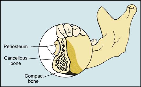
The periosteum (Latin for “surrounding the bone”) is the first layer of bone. It is a thin layer of whitish connective tissue and contains nerves and blood vessels. It supplies the cells from which the hard bone below the periosteum is built up. It is necessary for bone growth and repair, for nutrition, and for carrying away waste. It is the periosteum that is responsible for the life of the bone, and it is capable of repair. The inner layer is loose connective tissue that contains osteoblasts, or cells associated with bone formation. The periosteum is anchored to bone by Sharpey’s fibers, which penetrate the underlying bone matrix. Beneath the periosteum is dense, rigid compact bone.
Compact bone is hard, dense, and very strong (Fig. 7-3, B; see also Fig. 7-2). It forms the outer layer of bone, where it is needed for strength. This layer of bone is so dense that surgeons must use a saw or a bone bur instead of a knife to cut through it.
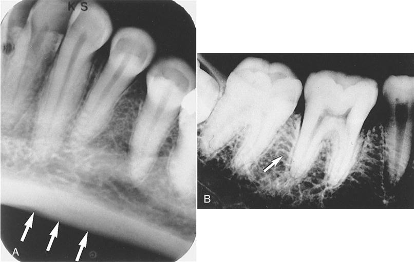
Cancellous bone is found inside the bone. It is lighter in weight and is not as strong as compact bone. Trabeculae (plural of trabecula) are bony spicules in cancellous bone that form a honeycomb pattern of spaces that are filled with bone marrow. On a radiograph, trabeculae appear as a weblike structure (see Fig. 7-3, A).
Bone marrow is a gelatinous material that produces white blood cells (which fight infection), red blood cells (which carry oxygen), or platelets (which help to stop bleeding).
Cartilage
Cartilage is strong but is more elastic than bone. It is found where bones join together. Cartilage is a tough, nonvascular (not associated with blood) connective tissue. In addition to its presence in the ends of bones, cartilage forms the nose and ears.

Joints
Joints, or articulations, are the areas where two bones come together (Fig. 7-4). The three basic types of joints are as follows:
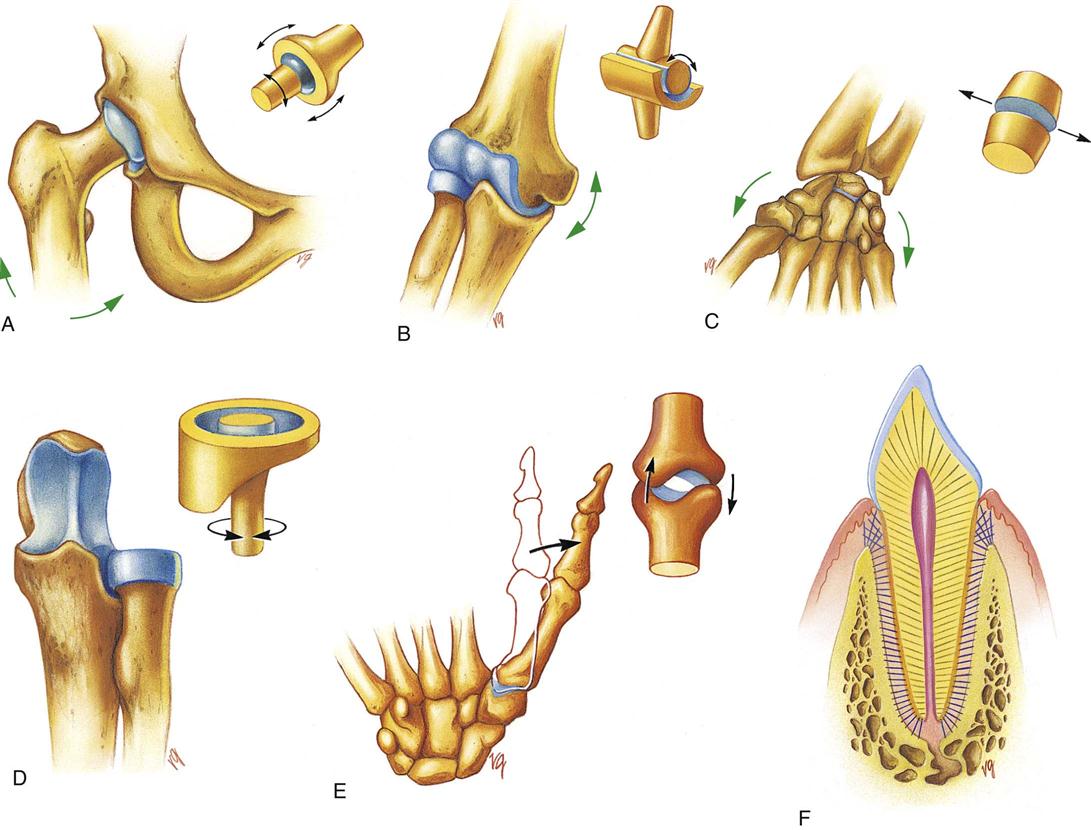
Ball-and-socket joints allow for movements such as flexion, extension, and limited rotation. Hinge joints allow movement in only one direction, similar to the hinge on a door. Gliding joints allow the bones to slide. The pivot joint at the base of the skull allows the head to rotate. The saddle joint allows movements such as touching the thumb to the fingers. Gomphoses are specialized joints that allow only very slight movement, such as attaching a tooth to the socket.

Muscular System
The muscular system gives us the ability to stand, walk, run, jump, move our eyes, smile, and frown (Figs. 7-5 and 7-6). For muscles to make the body move, they must work together. Each muscle consists of muscle tissue, connective tissue, nerve tissue, and vascular (blood) tissue. Many disorders can affect the muscular system (Table 7-3).
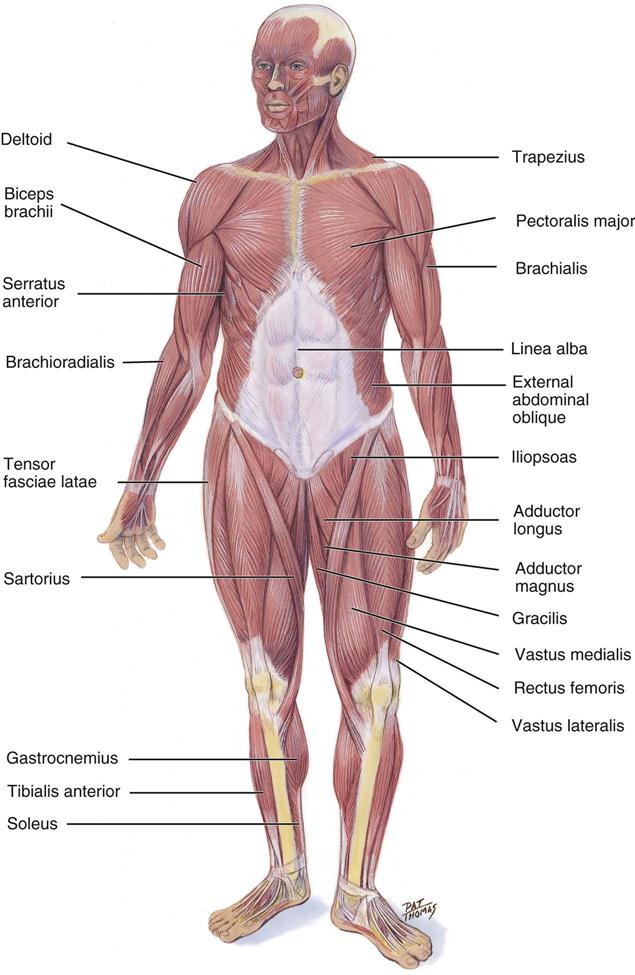
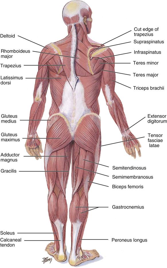
TABLE 7-3
Disorders of the Muscular System
| Disorder | Description | Signs and Symptoms |
| Contusions | Soft tissue trauma | Swelling, tenderness, and localized hemorrhage and bruising can restrict range of motion without producing loss of joint stability. |
| Strain | Injury of a muscle that has been stretched beyond its capacity | Small blood vessels around the area rupture, causing swelling in the area. The area becomes tender, and painful muscle spasms may occur. |
| Progressive muscular dystrophy | Includes nine types, all with unknown causes | Progressive muscle atrophy with organ involvement and weakness |
| Sprain | Injury to a joint that has been stretched beyond its normal range of motion, resulting in a tear | Depending on the severity of damage, it may include pain in the affected area, swelling, bruising, abnormal motion, and joint weakness. |
The muscular system is composed of more than 600 individual muscles. However, only three types of muscles have been identified: striated, smooth, and cardiac.
Striated Muscle
Striated muscles are so named because dark and light bands within the muscle fibers create a striped, or striated, appearance. Striated muscles are also known as skeletal, or voluntary, muscles. These muscles attach to the bones of the skeleton and make voluntary bodily motion possible. Voluntary muscles are so named because we have conscious (voluntary) control over these muscles. For example, you decide when to move your arms or legs.
Smooth Muscle
Smooth muscle fibers move the internal organs, such as the digestive tract, blood vessels, and secretory ducts leading from the glands. In contrast to the marked contraction and relaxation of striated muscles, smooth muscles produce relatively slow contraction.
Smooth muscles are also known as unstriated, involuntary, or visceral muscles. Unstriated muscles do not have the dark and light bands that produce the striped appearance seen in striated muscles. Involuntary muscles are so named because they are under the control of the autonomic nervous system and are not controlled voluntarily. For example, you do not decide when to begin digesting your lunch. Visceral muscles are so named because they are found in all the visceral (internal) organs except the heart. They are also found in hollow structures, such as the digestive and urinary tracts.
Cardiac Muscle
Cardiac muscle is striated in appearance but resembles smooth muscle in its action. Cardiac muscle forms most of the walls of the heart, and contraction of this muscle results in the heartbeat. Over a lifetime, the human heart beats 4 billion times and pumps 600,000 tons of blood. Physiologists think the reason why cardiac muscle is so durable is that it combines the power of striated voluntary muscles with the steady reliability of smooth, involuntary ones. Moreover, the cardiac fibers connect with each other and create a mutually supportive network of communications.

Muscle Function
Muscles are the only bodily tissues that have the ability to contract and relax. Contraction is the tightening of a muscle, during which it becomes shorter and thicker. Relaxation occurs when a muscle returns to its original form or shape. Muscles of the body are arranged in opposing pairs, so that when one muscle contracts, the other muscle relaxes. These opposing actions make motion possible.
Muscle origin is the place where the muscle begins (originates). This refers to the more fixed attachment, or the portion of the muscle that is toward the midline of the body.
Muscle insertio/>
Stay updated, free dental videos. Join our Telegram channel
VIDEdental - Online dental courses


