7: Cores

Chapter 7 Cores

Materials

Several dental materials can be used for core build-ups; each has different properties and therefore advantages and disadvantages. Enamel–dentine bonding using adhesive materials, such as composite resins or glass ionomer-based materials, allows a more conservative techniquecompared to amalgam which often requires additional tooth preparation to achieve adequate retention. Crown preparation can be carried out immediately following core build-up if the material used is command set by light curing. If the core is placed as a transitional restoration at an appointment prior to preparation then it should be sufficiently contoured to give occlusal stability, cause no food packing and maintain gingival health.

There are many materials on the market that are suitable for core build-up, but those based upon composite resin are increasing in use and many may say they are the most appropriate to use in contemporary practice. The use of amalgam has decreased in popularity, mainly because of health and environmental issues over its mercury content, its inability to bond to tooth structure and its colour. In a number of countries the use of dental amalgam has already been banned and this is only likely to extend to other countries in the future. However, it is still in use in sufficient countries and sufficient quantities to warrant its description in this textbook.

Composite resin (Figure 7.1)
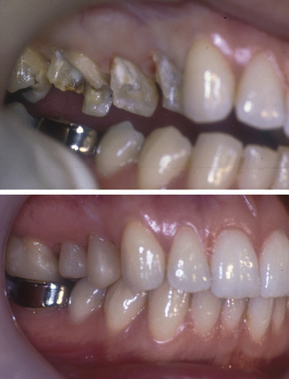
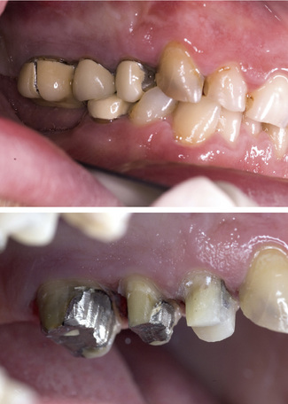
Composites consist of a resin, normally an aromatic dimethacrylate such as BisGMA, and filler particles such as quartz, silica and other types of glass. The type, particle size and content of the filler particles control the properties of the material. Barium- or strontium-containing glasses are also added to make the material radio-opaque. Many composites have a variation in particle size. So-called hybrid composites contain large filler particles (15–20 µm) and smaller colloidal silica (0.01–0.05 µm) particles. Microfilled composites (average filler particle size = 0.02 µm) can be highly polished, but this is a property obviously not necessary for a core material. The most effective core materials are hybrid composites and more recently the newly developed low-shrink composites (e.g. Filtek LS Low Shrink Composite (3M ESPE) based on silorane chemistry) might prove to be effective. Composites have a compressive strength similar to dentine and higher tensile and flexural strengths. They can be bonded to tooth structure when used with a dentine-bonding agent; however, placement is technique sensitive.

When using composites it is important to prevent moisture contamination with saliva or blood and, therefore, where possible, rubber dam isolation is recommended. Visible light cured composites should be placed in increments to reduce problems with shrinkage on setting. Polymerization contraction may increase the risk of marginal leakage and post-treatment sensitivity. The lower shrink composites that are being introduced may overcome this problem in the future but will need assessing as the materials develop. Water sorption occurs with composites and can lead to expansion of the material. This, however, takes some time to occur and is unlikely to have an impact on the provision of indirect restorations. Any dimensionalchange that does occur will be easily compensated for by the placement of die relief on the master die in the laboratory. Composite is the material of choice for a core when an all-ceramic crown is planned (Figure 7.1).

Newer hybrid composite core materials are available with various additives such as fibres, ceramic fillers, titanium and lanthanide, that claim to improve the mechanical properties of the material. Examples of these are Paracore (Coltène Whaledent), Ti-core (Essential Dental Systems, Inc.), Light-Core (Bisco, Inc.), Coradent (Vivadent) and Core Paste XP (Den-Mat). Composite core materials are available with a blue pigment to allow the clinician to distinguish between tooth structure and material during preparation (e.g. MultiCore Flow Blue, Ivoclar). Some composites are also packable, facilitating contact point formation and reducing the risk of voids.

Resin-modified glass ionomers (RMGIs)

In the early 1990s a water-soluble resin (hydroxyethyl methacrylate, HEMA) was added to conventional GIC to improve the physical properties, as such the material cures by an acid–base reaction (the glass ionomer component) and a resin polymerization, which is either chemical or light activated or both (dual cured). Some RMGIs are specifically advocated as core materials such as Vitremer core build-up material (3M ESPE). Whilst most of these materials are tooth coloured, their use beneath all-ceramic restorations should be avoided as they undergo hygroscopic expansion which could lead to ceramic fracture. Resin-modified glass ionomer is an ideal core material for metal or metal–ceramic crowns, inlays, onlays or bridges, or reinforced core ceramics; however, like chemically cured glass ionomers, it needs significant remaining tooth structure to be effective.

Amalgam (Figure 7.2)

Dental amalgam consists of mercury combined with a powdered silver–tin alloy with the addition of copper, palladium and other elements. High copper amalgam alloys have a copper content of 30% and on setting have a smaller concentration of the gamma-2 phase, which means they are less easily deformed, stronger in compression, and have reduced potential for corrosion. The alloy particles can be spherical, lathe cut or admixed. Amalgam can be the material of choice for core build-ups on posterior teeth and is still used by some practitioners. Its use is, however, on the decline and is only likely to decline further in the future. Unfortunately, as the material takes 24 hours to reach maximum compressive strength, crown preparation has to be carried out at a second appointment. The spherical, high copper alloys reach their maximum strength faster and can be prepared 10–15 minutes following placement rather than the normal 24 hours; however, even this is a long time to wait in a busy practice. As amalgam does not adhere to tooth structure it has to be mechanically retained or chemically bonded. It performs well if properly condensed and is not contaminated by large amounts of blood or saliva during placement.

Vital teeth

Methods of core retention for vital teeth

There are no absolute guidelines, but if more than 50% of tooth structure has been lost or removed, additional methods of retention are usually required. Retention can be either mechanical, when using amalgam, or adhesive. The latter is the most common as composite is the preferred material for a core.

Adhesive retention

Composite resins and dentine bonding

Bonding of composite to etched enamel has made a huge advance in operative dentistry, allowing aesthetic restorations to be placed and clinical techniques such as minimum preparation (resin retained) bridges. Bonding of hydrophobic composite resins to physiologically wet dentine has also made a massive leap forward over the last two decades. Generally the dentine surface is chemically treated with an acid to allow the mechanical interlocking of resin around the dentinal collagen, producing a layer known as the hybrid layer. The bonding agent, which is normally an unfilled resin, is then applied to the dentine surface and light cured. It co-polymerizes with the resin already present in the collagen matrix, locking it onto the dentine and providing a more suitable surface for bonding with resin composite materials.

The steps involved in dentine bonding have changed over the years. Initially a separate etch, primer and bond were used. With this technique there was a risk that during drying the collagen may collapse and ruin formation of the hybrid layer. Two-step systems have either a separate etch with the prime and bond combined in one bottle or the etch and prime combined with a separate bond. The latter has the advantage that the self-etching priming agent does not have to be washed off the dentine. One-step bonding systems are similar to self-etching priming materials but have the bonding agent added also. They have been shown not to etch the enamel as effectively as phosphoric acid.

Stay updated, free dental videos. Join our Telegram channel

Jan 9, 2015 | Posted by in Operative Dentistry | Comments Off on 7: Cores

VIDEdental - Online dental courses

Get VIDEdental app for watching clinical videos