6 Methods of Extraction
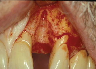
Implant dentistry has not only revolutionized the ability to treat fully and partially edentulous patients; it has also enlightened the clinician with the virtues of bone preservation following tooth extraction. The ability to treat a site with a dental implant begins with one’s decision on the method of tooth extraction. This is of critical importance, especially when an immediate implant placement protocol is desired, given that this modality of treatment demands that the implant be primarily stable upon insertion via the lateral walls of the socket or virgin bone available at the apex (Lazzara 1989). Quite often, iatrogenic trauma to the surrounding hard tissues of the socket may prevent primary stability from being obtained and the immediate procedure will need to be aborted, with an early or delayed approach to placement required (Figure 6.1) (Chen, Wilson, et al. 2004).
Despite various claims of certain extraction techniques as being “atraumatic,” all extraction methods impose some level of hard and/or soft tissue trauma. Every tooth one encounters for extraction offers unique challenges when immediate implant placement is to follow (Fugazzotto 2002; Fugazzotto 2006; Fugazzotto 2008). This chapter provides insight into the various methods, instruments, and techniques available to the clinician to best preserve the hard and soft tissues, in the face of the unique challenges each extraction site presents.
Figure 6.1 Buccal view of iatrogenic trauma with tooth extraction.
DENTAL FORCEPS
It is probably correct to assume that the extraction of teeth, due to caries or fracture, dates back to the beginning of mankind. The use of an extraction forceps was described by Aristotle (384–322 BC) as two levers acting in contrary sense with a single fulcrum (Ring 1985). Forceps today have evolved to a variety of shapes and sizes, often specific to both deciduous and permanent teeth in the maxilla or mandible (Figure 6.2). These devices are designed to grip the affected tooth by the anatomic crown, remaining root structure, or furcation region, and provide the clinician with a greater mechanical advantage to remove the tooth. One must keep in mind the tooth is maintained in the socket via the periodontal ligament. Therefore, the objective in the extraction of teeth is to separate or sever the periodontal ligament from the tooth, most often using a rotation movement applied to the long axis of the root (Leonard 2002). Quite often, practitioners view the purpose of the dental forceps as to enable the rocking of the tooth in a buccal-lingual direction in an effort to expand the alveolar process for the delivery of the tooth.
When this protocol is followed, it becomes a contest between the integrity of the root of the tooth and the integrity of the buccal bone plate. If the bone is stronger than the root, then the root fractures. If the root is stronger than the bone, then a resulting dehiscence will arise with the fractured buccal plate. In light of the desire to preserve the intact alveolus for the purpose of immediate implant placement, the forceps’ role should be limited to assisting in the stretching and severing of the periodontal ligament, and to delivery of the tooth or root. This implies that all teeth should be treated as single roots and therefore multi-rooted teeth should be sectioned prior to applying the site-appropriate forceps (Figures 6.3 and 6.4). When used correctly, the forceps should generate a gradual force applied clockwise to the long axis of the root for approximately 10 seconds before reversing the force counterclockwise. In many instances, it will become necessary to narrow the coronal aspect of the crown in a mesial-distal direction to permit proper rotation. This exercise is repeated until the tooth loosens and is then delivered coronally to prevent fracture of the labial plate. Care should be given to observe any minute fractures of the thin buccal plate when rotating the tooth. Should this occur due to an oval-shaped root, a consideration should be made to section the root vertically using a surgical bur and handpiece to avoid further trauma.
Figure 6.2 Illustration of anatomical forceps for a maxillary incisor.
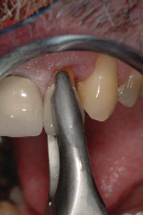
Figure 6.3 Buccal pre-op view of tooth #19.
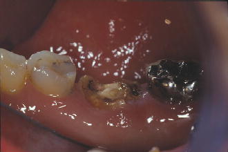
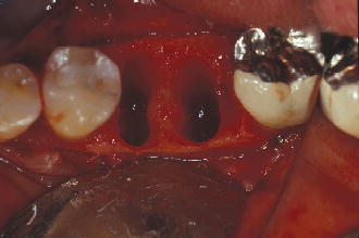
Figure 6.4 Occlusal view of #19 site following sectioning and extraction.
DENTAL ELEVATORS
A simple machine is a device designed to apply or transmit force or torque. Simple machines consist of levers, inclined planes, wheels, screws, and pulleys. A dental elevator is therefore a combination of two simple machines—a lever and an inclined plane. The principle of a lever was originally described by Archimedes (circa 350 BC). The first reference to the use of a simple lever (elevator) to lift a tooth from its socket was by Abulkasim (1050–1122 AD) (Atkinson 2002). Much like dental forceps, elevators are available in a variety of shapes and sizes to apply a vertical or lateral force to a tooth root (Figure 6.5). When used with a vertical force, the wedge shape of an elevator provides the clinician a greater mechanical advantage to initiate the luxation of a tooth for its removal when pushed along the long axis of the root surface (Misch 2008). At minimum, the width of dental elevators measures 1.5 mm, while the space of the periodontal ligament ranges from 0.1 mm to 0.3 mm. Using an elevator for the purposes of initially luxating a tooth along its long axis most frequently results in crushing the thin interproximal bone along the mesial and/or distal, and in the esthetic zone, may affect the ability to develop an ideal papilla form. Another concern when using elevators to luxate teeth along the long axis is the possibility of increasing the mesial distal width of the socket, preventing an immediate implant from engaging the lateral walls of the />
Stay updated, free dental videos. Join our Telegram channel
VIDEdental - Online dental courses


