Variations in Transition of Maxillary Incisors*
The morphology of the maxilla deviates markedly from that of the mandible. The maxilla is connected with other bones of the skull by sutures and does not act as a separate entity, as is the case for the mandible. Its shape and structure differ accordingly. Further, the maxilla can increase in size in the median plane because the intermaxillary suture maintains its growth potential until the development of the dentition is completed.
The space available within the maxilla for the permanent incisors before emergence is more limited than in the mandible. The amount by which maxillary permanent incisors exceed their predecessors in crown dimensions is greater than that for mandibular incisors. The crowns of maxillary permanent incisors are oriented within the jaw in a typical way; before emergence the spatial conditions in the maxilla allow a larger variation in the arrangement both vertically and sagittally of the permanent incisors than in the mandible. Consequently, the eruption process of maxillary incisors is characterized by a larger variation than that of mandibular incisors.
The transition of maxillary incisors is discussed in relation to the size and shape of the anterior section of the apical area. The variation in location of permanent incisors and canines before emergence and their relation to the adjacent deciduous predecessors are indicated. Eruption is treated in preemergence and postemergence phases. Five eruption patterns are presented by means of longitudinal records of dental casts and schematic drawings.
5.2 The anterior section of the apical area and the arrangement of anterior teeth before emergence
The location of the anterior section of the apical area in the maxilla is mainly determined by the size, shape, and anteroposterior orientation of the piriform aperture. A large variation exists in these respects. The forming canines are located in close proximity to the piriform aperture. As a result, the transverse distance between the two forming permanent canines can differ and their anteroposterior location can vary considerably. The central permanent incisors are formed in close proximity to the floor of the anterior part of the nose and with the intermaxillary suture in between. The morphology of the bony structure of the anterior part of the maxilla adjacent to the nose determines the space available in transverse and anteroposterior directions for the forming parts of the permanent incisors and canines and also the range available for location of the apices of these teeth upon complete eruption.
The anterior section of the apical area is bounded laterally by the location of the permanent canines and sagittally by the labial and palatal walls of the alveolar process. The space available for the roots of the deciduous teeth and crowns of the permanent teeth prior to their emergence is limited. Maxillary permanent central incisors have the widest crowns in relation to their roots. The anterior section of the apical area must accommodate these crowns together with the roots of their predecessors in a compartment that later will serve to contain the roots of the permanent incisors only. No other region of both jaws demonstrates such a comparatively large difference in space required in early and late stages of development of the dentition.
The maxillary permanent canine starts its formation lateral to the piriform aperture and at the largest distance from the occlusal plane. The permanent central incisor is formed near the midline and more anteriorly than the lateral incisor. The lateral incisor starts its formation at a more inferior level than the central edge incisor, consequently its incisal edge is located closer to the occlusal plane than the incisal edge of the central incisor, until the latter starts to erupt.
The cusp tip of the permanent canine and the cementoenamel junction of the adjacent lateral permanent incisor are more or less at the same level. However, some variation exists, depending on the differences in height of formation of the permanent incisors and canine and on the crown length of the lateral incisor.
The lateral permanent incisor is located palatal to the central incisor and canine. The permanent maxillary central incisor overlaps the lateral incisor (the amount of overlap depending on the space available for the lateral incisor, as determined by the intercanine distance, and on the crown size of the central and lateral incisors).
Before the maxillary permanent incisors emerge they show less frequent rotations than do mandibular incisors. This is probably related to differences in morphology between the anterior regions of the maxilla and mandible and differences in size and shape between the maxillary and mandibular permanent incisors. On the other hand, the maxillary permanent incisors with their relatively large mesiodistal crown dimensions show a larger variation when rotated than do mandibular incisors.
The relation between the space available and the space needed for arrangement of the tooth material within the anterior section of the apical area before the permanent incisors emerge shows a marked variation (Figs. 5-1 to 5-3). The anterior section of the apical area is of adequate size when the distance between the maxillary permanent canine crowns is large in relation to the mesiodistal crown dimensions of the maxillary permanent incisors. Frequently, a large anterior section of the apical area is associated with large diastemata in the deciduous dentition. There is no initial contact between the distal surface of the central permanent incisor crown and the root of the adjacent lateral deciduous incisor. Consequently, the distance between the maxillary deciduous canines will not increase or will only slightly increase before and during emergence of the maxillary central permanent incisors. However, large diastemata in the maxillary anterior region are not always associated with a large anterior section of the apical area, and no or small diastemata are not always associated with a small anterior section. As for the mandible, only a limited correlation exists between the mesiodistal crown dimension of the maxillary incisors of the two dentitions.
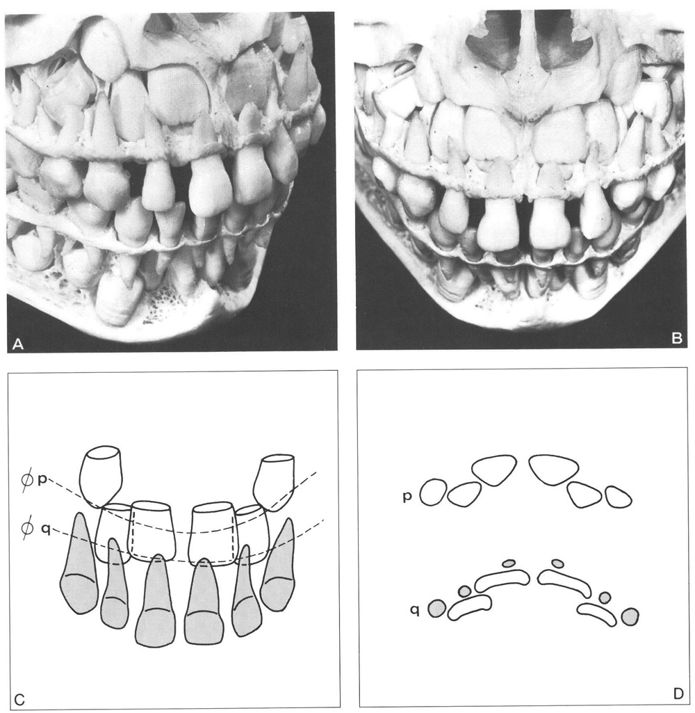
Fig. 5-1 Large anterior section of the apical area in the maxilla.
A, B Large diastemata between the crowns of the anterior deciduous teeth. The width of the piriform aperture and the associated distance between the crowns of both maxillary permanent canines are large. The permanent incisors show a more or less perpendicular mesiodistal angulation in relation to the occlusal plane. The crowns are not rotated. The central permanent incisor only slightly overlaps its neighbouring lateral incisor. Note the more caudal position of the incisal edge of the lateral permanent incisor than that of the central incisor.
C Schematic drawing of B with dotted lines indicating the sections presented in D. The distal surfaces of the crowns of the maxillary central permanent incisors do not contact the roots of the adjacent lateral deciduous incisors.
D In the region of the incisal borders of the permanent incisors (q) the lateral incisors are palatal to the central incisors. Note the relations between the roots of the deciduous teeth and the crowns of the permanent teeth. In the region of the cementoenamel junctions of the permanent incisors (p) the lateral incisors are also palatal to the central incisors. Note the triangular shape of the cross section of the roots of the permanent incisors.
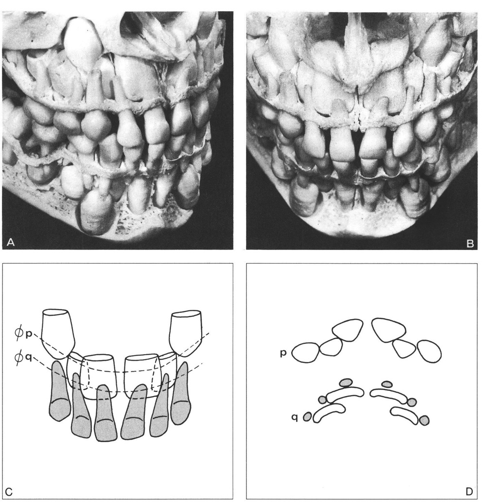
Figure 5-2
Fig. 5-2 Medium anterior section of the apical area in the maxilla.
A, B The diastemata between the anterior deciduous teeth are medium-sized. The width of the piriform aperture and the associated distance between the crowns of both maxillary permanent canines are also medium-sized. The maxillary central permanent incisors are almost perpendicularly angulated in relation to the occlusal plane; they are not rotated. The permanent canine crown slightly overlaps the lateral incisor.
C The distal corners of the central permanent incisors contact the mesial surfaces of the roots of the adjacent lateral deciduous incisors.
D In the region of the incisal borders of the permanent incisors (q) a close proximity exists between the crowns of the permanent teeth and the roots of the deciduous teeth. Note the contacts described under C. In the region of the cementoenamel junctions of the permanent incisors (p) the teeth overlap. Note the close proximity between the permanent canines and the lateral permanent incisors.
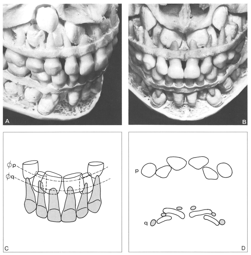
Fig. 5-3 Small anterior section of the apical area in the maxilla.
A, B No diastemata between the deciduous incisors. The width of the piriform aperture and associated distance between both maxillary canine crowns is small. The distally angulated central permanent incisors are close together just below the floor of the nose. The close proximity between the formation area of a maxillary central permanent incisor and the median plane produced distally angulated crowns that are also mesiopalatally rotated. The central permanent incisors overlap the lateral incisors by approximately half their crown width. The permanent canine crown overlaps approximately one half the adjacent lateral permanent incisor.
C The distal corners of the crowns of the central permanent incisors contact the mesiobuccal surfaces of the roots of the adjacent lateral deciduous incisors.
D In the region of the incisal borders of the permanent incisors (q) the mesial incisal corners of the central incisors are abnormally palatally positioned. The contact between their distal incisal corners and the roots of the adjacent lateral deciduous incisors is not favourable. Close proximity exists between the central incisors in the region of the cementoenamel junctions of the permanent incisors (p). The space available for the arrangement of the permanent teeth is small.
The transition of maxillary and mandibular incisors is illustrated and explained in Fig. 2-4. Figure 5-4 shows six prepared skulls illustrating the transition of the maxillary incisors. This figure further shows the large variation in transition of maxillary incisors together with typical abnormalities.
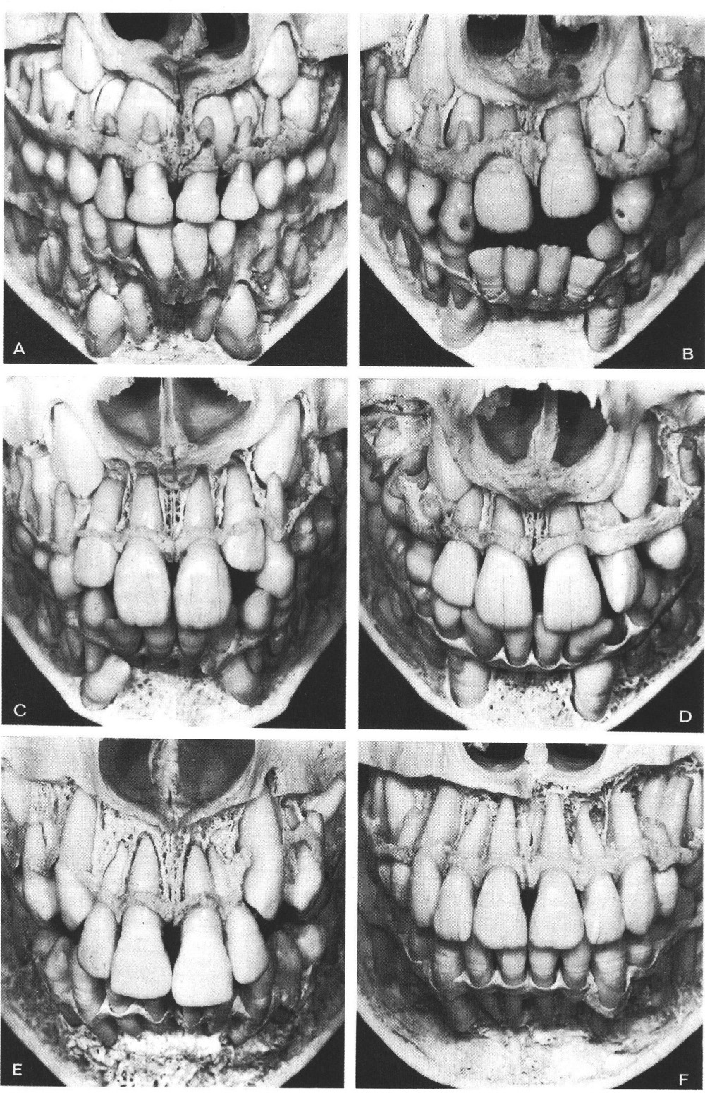
Figure 5-4
Fig. 5-4 Six preparations of human skulls with small and medium anterior sections of the apical area in the maxilla that demonstrate different aspects of and disturbances in the transition of the maxillary incisors and canines.
A Small anterior section of the apical area with all maxillary deciduous teeth still present. The mandibular central permanent incisors have already reached the level of the occlusal plane. The maxillary central permanent incisors have started their eruption and passed the adjacent lateral incisors. Note the contact between the distal corners of the crowns of the central permanent incisors and the lateral deciduous incisor roots. The asymmetry in the location of the maxillary central permanent incisors shows up in the morphology of the floor of the nose. No diastemata or only small diastemata are present between the anterior deciduous teeth. The width of the piriform aperture and the distance between the maxillary permanent canines are small. The central permanent incisors overlap approximately half the width of the crown of the adjacent lateral incisors.
B Small anterior section of the apical area, asymmetrically erupting central permanent incisors, and premature loss of the maxillary left lateral deciduous incisor. In the maxillary right quadrant contact exists between the central permanent incisor, the lateral deciduous incisor, and the deciduous canine. The distal and lateral movement of the deciduous canine, associated with the eruption of the central permanent incisor, can only take place on the right side and not the left. The maxillary lateral left permanent incisor has an abnormally shaped crown and is located in a rotated position.
C Medium anterior section of the apical area; maxillary central permanent incisors fully erupted and the lateral incisors partially erupted. The lateral incisors started to erupt and could move labially after the central incisors had descended. Note the difference in labial inclination between the central and the lateral incisors. The space between the root of the lateral incisor and the permanent canine crown is larger on the left than on the right side. The left lateral incisor will have more freedom to change in (mesiodistal) angulation during eruption than the right one.
D Small anterior section of the apical area with all four maxillary permanent incisors fully erupted. The maxillary left deciduous canine has been prematurely resorbed in association with the eruption of the adjacent lateral permanent incisor. The latter attains a more distal position in the dental arch than it otherwise would have done. Note the deviation in the midline of both dental arches.
E Medium anterior section of the apical area. The central and lateral incisors are fully erupted and their roots completely formed. The right permanent canine has emerged and erupted into contact with the crown of the lateral incisor. On the left side the canine has not erupted so far; the contact with the lateral incisor is not yet established. The roots of the central and lateral incisors are still close together. After the eruption of the canines the roots of the lateral incisors will be able to move distally.
F Medium anterior section of the apical area with fully erupted canines. The formation of their roots, however, is not yet completed. Some space exists between the roots of the lateral and the central incisors. Note the differences in labiolingual inclination between the central and lateral incisors. The apices of the lateral incisors are more palatally located than those of the central incisors. This difference is in accordance with the original sites of tooth formation and their subsequent position before eruption.
In general, a central permanent incisor starts to erupt in the same direction as its orientation within the jaw. The descending central incisor passes the adjacent lateral incisor and the amount of overlap gradually reduces. As a rule, the overlapping has been fully eliminated before complete eruption of the central incisor, when its cementoenamel junction has reached the level of the incisal corner of the lateral incisor. Sufficient space is then available for the maxillary lateral permanent incisor to descend. In accordance with its original location within the jaw, the lateral incisor will then erupt in a more labial direction than the central incisor. When a permanent incisor erupts in a small anterior section of the apical area, not only the preceding deciduous tooth but also the adjacent deciduous tooth may become resorbed. A lateral deciduous incisor may be lost in association with the eruption of a permanent central incisor; a maxillary deciduous canine may prematurely exfoliate when the adjacent permanent lateral incisor erupts. The premature loss of one or two central deciduous incisors due to trauma (Fig. 5-5) will not result in disturbances in the development of the dentition, if the position and formation of their successors have not been affected.101
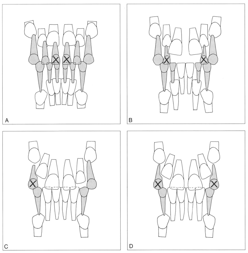
Fig. 5-5 Premature loss of deciduous incisors and canines in the maxilla due to trauma (A) or in association with the eruption of adjacent permanent teeth in a small anterior section of the apical area (C, D, E). (From Van der Linden.223)
A Premature loss of one or two maxillary central deciduous incisors due to trauma will not disturb development of the dentition when the position and formation of the successors are not affected.
B Premature loss of both lateral deciduous incisors due to resorption associated with the eruption of the permanent incisors. The increase in intercanine width, which is normally associated with the eruption of the maxillary central permanent incisors and is based on the contact between the crowns of the central permanent incisors and roots of the adjacent lateral deciduous incisors, will not take place.
C Premature loss of the maxillary right deciduous canine associated with the eruption of a large lateral permanent incisor. The permanent incisors will subsequently migrate toward the open space.
D Premature loss of both maxillary deciduous canines associated with the eruption of the lateral permanent incisors.
5.4 Eruption after emergence and subsequent change in orientation
The position of the maxillary permanent incisors becomes influenced after emergence by forces of the lips, tongue, and sometimes by factors of an abnormal nature, for example thumb-sucking. The maxillary central permanent incisors do not emerge lingual to the position originally occupied by their predecessors, as is the case in the mandible. By nature of their labial inclination they soon start to protrude labially beyond the places where previously the labial surfaces of their predecessors were located. Contact is then established with the upper lip, and under its influence the lateral inclination becomes reduced. Upon further eruption the incisors reach the lower lip and establish contact with the mandibular incisors (Fig. 5-6). The position attained upon complete eruption depends on the original position and orientation of these teeth within the jaw and the eruption path as it becomes influenced after emergence by the factors mentioned above. Local conditions within the dental arches also have an influence. Crowding will be demonstrated by rotation, overlapping, eversion, linguoversion, inversion, or a combination of these. A sagittal deviation in the relation between the dental arches will affect the ver/>
Stay updated, free dental videos. Join our Telegram channel

VIDEdental - Online dental courses


