CHAPTER 5 Systematic Pretreatment Evaluation of the Edentulous Maxilla
Three factors are key determinants for the successful treatment of the completely edentulous maxilla with a fixed restoration. These factors, which can be determined early in the examination process, are (1) the presence or absence of a composite defect; (2) the visibility or lack thereof of the residual ridge crest without the denture in place, with normal smile and without use of retractors; and (3) the amount of bone available in three separate zones of the maxilla, as revealed in a panoramic survey. Prior to discussing each of the three factors for the systematic evaluation of the edentulous maxilla, it may be prudent to establish several baseline criteria. The implant-support requirements of all three fixed restorative options discussed in this chapter are the same as the requirements for a fully implant-supported, nonresilient maxillary restoration. A minimum of four implants should be used, although the option to place more than four may be considered, depending on the available bone volume and other functional considerations.1,2 The most important factor is the arch-form distribution of those implants with both posterior and anterior support. Evaluation of the three radiographic zones allows for a preoperative determination of whether adequate arch-form support for a fixed restoration is achievable to support the planned occlusal plane. As a general principle, cantilevers in fixed maxillary restorations should be avoided or minimized to one tooth to achieve an adequate functional occlusion.3–7 Several descriptions of fixed prostheses used to restore completely edentulous maxilla have been published.8–11 This discussion focuses on the application of three principal designs for implant-supported dental prostheses. These three variations have been chosen based on their ability to restore a broad range of soft tissue deficits. They are (1) metal-ceramic restoration, (2) fixed-hybrid restoration, and (3) fixed-removable restoration.
Fixed-Maxillary Prostheses
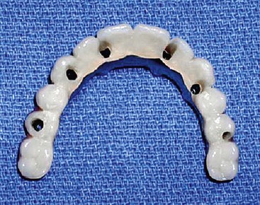
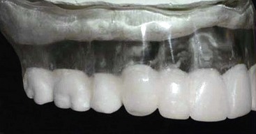
Metal-ceramic restorations may be either screw- or cement-retained.12–14 Recognizing that ceramic restorations can include teeth that are longer than normal and gingival replacement, emphasis is on metal-ceramic restorations used to replace the clinical crowns of missing teeth only (Figure 5-1).
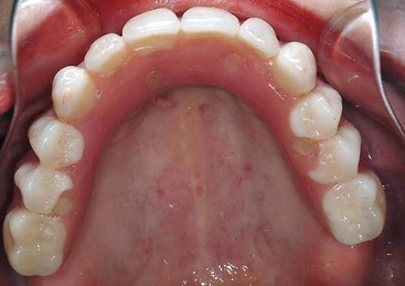
The hybrid prosthesis is a denture tooth and acrylic design with either a milled titanium or cast-gold framework (Figure 5-2). Practitioners using early designs of implant-supported denture tooth and acrylic fixed prostheses reported phonetic changes as a routine complication, caused by air escaping during speech.15 A later design known as profile prosthesis16 uses a framework design with subgingival abutment emergence that allows an acrylic resin wrap; the wrap butts up against the tissue as an ovate pontic so that air does not escape and cause phonetic problems. Because a ridge lap is avoided with the convex emergence from the ridge crest, oral hygiene access can be maintained in a manner similar to natural-tooth, fixed, partial-denture pontics.16 A variation of this design uses gingival porcelains or composite with all-ceramic crowns cemented to the framework, which can be used if a porcelain restoration is desired.
For situations in which a labial flange is desirable, a fixed-removable prosthesis can be made with any number of attachments. Figure 5-3 demonstrates a fixed-removable design known as a Marius bridge that is nonresilient and fully implant-supported.17 Fixed-removable designs use a milled titanium or cast meso bar supporting a patient-removable superstructure held in place with a locking mechanism. This allows a ridge lap or flange design, with a suprastructure removable for oral hygiene access. Because a fixed-detachable restoration does not depend on soft tissue support, no unnatural palatal extensions are required.
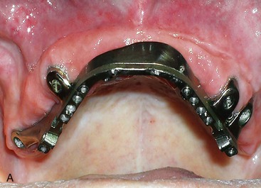
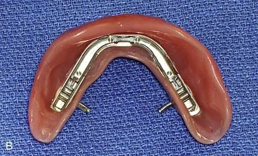
FIGURE 5-3 The Marius bridge, a fixed, detachable prosthesis, may be fabricated for full-arch replacement.
To determine which of these prostheses is most appropriate, identification of the presence or the lack of a composite defect and the relationship of the transition line to the patient’s smile line needs to be made. Evaluation of these factors is not intended to be a substitute for thorough diagnosis and development of a treatment plan. However, such evaluation can provide differential diagnosis information specific to the aesthetic, prosthetic, and biomechanical requirements of fixed, implant-supported maxillary restorations.18
Presence or Absence of a Composite Defect
The first criterion to consider is the presence or absence of a composite defect. Edentulous patients may present with intact alveolar bone volume and be missing only the clinical crowns, or they may present with missing teeth as well as resorption of their alveolar bone and loss of soft tissues. Differentiating between these two types of patients is essential to creating an aesthetic, definitive fixed prosthesis. Patients missing soft tissue and underlying supporting bone in addition to teeth have a composite defect (Figure 5-4). To evaluate the relative soft and hard tissue deficiencies, it is advisable to use a denture or denture set up in wax in which proper tooth position and interarch relationship (proper vertical dimension of occlusion) has been confirmed.
With a satisfactory denture, thickness of the maxillary denture base and flange can be used to assess the presence or absence of composite defect. A thick denture base and flange indicate moderate to advanced resorption of the maxilla. In situations in which minimal soft and hard tissue resorption has occurred, a “tooth-only defect” is present. For this group of patients, a thin denture base and a very thin or absent flange, especially in the anterior sextant, indicate an intact alveolus19 and therefore the absence of composite defect.
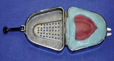
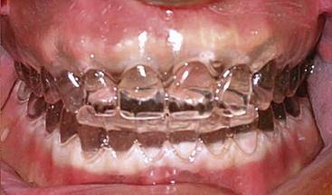
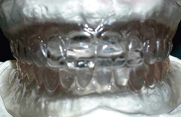
It should be noted that defects resulting from resorption of bone and missing soft tissue occur in both the horizontal and vertical planes, and may not be immediately obvious. To fully assess the presence or absence of a composite defect, duplication of the confirmed denture or tooth set-up by the dental technician or dentist using a denture duplicator can be useful (Figure 5-5). A transparent acrylic resin duplicate of the patient’s denture is then placed intraorally, and the position of the cervical portion of the teeth and their relationship to the crest of the edentulous ridge is noted. For patients who present with no space between the cervical portion of the duplicated denture teeth and the edentulous ridge in either horizontal or vertical planes, a tooth-only defect is diagnosed (Figure 5-6). In this situation, interarch space minimum requirements for the implant system and desired restoration still need to be observed. For patients who present with moderate to significant space between the cervical portion of the duplicated denture teeth and the edentulous ridge, a composite defect is identified (Figure 5-7). Table 5-1 illustrates these considerations.
| Intraoral Status | Diagnosis | Definitive Prosthesis |
|---|---|---|
| No space between the cervical portion of the duplicate denture teeth and the edentulous ridge | Tooth-only defect | Metal-ceramic |
| Moderate to significant space between the cervical portion of the duplicate denture teeth and the edentulous ridge | Composite defect | Marius bridge (fixed-detachable) or profile prosthesis (hybrid) |
Stay updated, free dental videos. Join our Telegram channel
VIDEdental - Online dental courses


