5
Radiographic Analysis
Periapical Survey
Periapical radiographs give useful information about caries, periodontal condition, periapical pathology, shape of the roots, size of the teeth, position of impacted teeth, and spatial location of teeth not yet erupted. Measurements of the mesial-distal widths of the periapical images of nonerupted premolars and upper canines are essential for the prediction of tooth size in the Hixon-Oldfather and Iowa mixed-dentition space analyses. A periapical survey of a patient in the early mixed dentition is illustrated in Figure 5.1. These radiographs give an accurate image of the roots that serve as a pretreatment baseline for the posttreatment assessment of root resorption. The pretreatment radiographs can also show the presence of root resorption before treatment. A 16-inch-long cone paralleling or right angle technique is recommended for taking the periapical radiograph.
Figure 5.1. Periapical survey of the early mixed dentition. Arrows point to the tooth images measured for the revised Hixon-Oldfather and Iowa tooth size prediction methods.
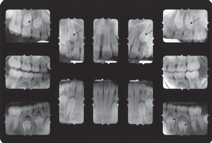
During treatment, periapical radiographs are used to monitor the position and movement of nonerupted teeth and the growth of the roots of developing teeth. At the end of active treatment, these radiographs can assess the presence and effect of root resorption.
Panoramic Radiograph
The panoramic radiograph gives a complete view of the dentition and supporting bones (White and Pharoah 2004). The stage of development of nonerupted teeth and the dental age of the patient can be determined by rating root development of several teeth. The shape of the condyles of the mandible can be observed, and abnormal or asymmetric shapes of the condyles can be noted and related to patient symptoms. Views of the relationship of nonerupted third molars to second molars and surrounding structures can help shape treatment planning decisions for these teeth.
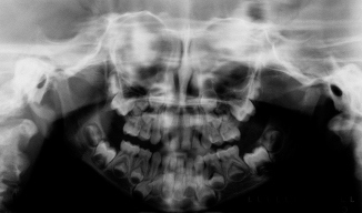
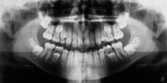
Several panoramic radiographs illustrate different developmental stages of growth, ankylosis, congenitally missing teeth, and impacted teeth in Figures 5.2 through 5.12. Figure 5.2 shows the erupted primary dentition and all of the developing nonerupted permanent teeth, except for the third molars. The early mixed dentition is shown in Figure 5.3. At this stage of development, the permanent incisors and first molars are erupted. In the late mixed dentition, at least one of the permanent canines or premolars has erupted. Figure 5.4 shows a patient with erupted permanent incisors, canines, first premolars, and first molars. The third molar buds are visible at this stage of development. The permanent teeth of an adult female are shown in Figure 5.5. All the permanent teeth, including the third molars, are in occlusion.
Figure 5.3. Panoramic of the early mixed dentition.
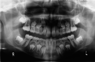
Figure 5.5. Panoramic of an adult dentition including third molars.
(Courtesy of Dr. Harold Bigelow.)
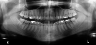
Figure 5.6. Panoramic showing ankylosed tooth T and tipping of tooth #30 that have prevented the eruption of tooth #29.
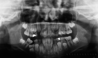
Figure 5.7. Panoramic of a male patient with aberrant second premolars and taurodont molars.
(Courtesy of Dr. Cynthia Christensen.)
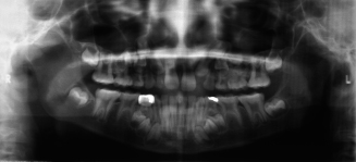
Figure 5.8. Panoramic of a patient congenitally missing an upper right second premolar and both lower second premolars. Primary second molars are retained and ankylosed (arrows).
(Courtesy of Dr. Theresa Juhlin.)
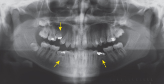
Figure 5.9. Panoramic of a patient who lost tooth A prematurely, which allowed the mesial migration of the upper right first molar that impacted the upper right second premolar.
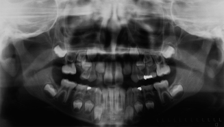
Figure 5.10. Panoramic of a patient who lost tooth K prematurely, which allowed the mesial migration of the lower left first molar that impacted the lower left second premolar.
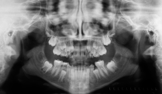
Figure 5.11. Panoramic of a patient with both maxillary canines impacted on the palatal side of the arch. The upper primary canines are retained.
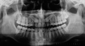
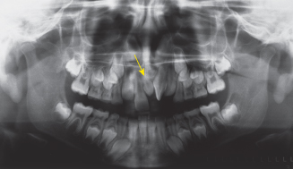
Figure 5.12. Panoramic of an early mixed dentition with a supernumerary tooth (mesiodens) located between the maxillary central incisors (arrow).
(Courtesy of Dr. Samir Bishara.)
The panoramic radiograph of a patient in the early mixed dentition who had an ankylosed mandibular right primary second molar is shown in Figure 5.6. The ankylosed molar sank below the normal teeth on either side of it, as growth of the alveolar bone took the normal teeth farther vertically. By the time a pediatric dentist saw the patient, the mandibular right permanent first molar had tipped forward over the occlusal surface of the akylosed primary molar. The mandibular right permanent second premolar is impacted beneath the ankylosed primary molar. Figure 5.7 is a panoramic radiograph of an early mixed-dentition patient (male) whose developing second premolars are displaced mesial to his primary second molars. Also, all of his erupted primary and permanent molars are prismatic or taurodont. In taurodont teeth, the pulp chamber is elongated and the distance between the bifurcation of the roots and the cementoenamel junction is greater than normal (Kovacs 1971). Normal distances are 4.8 ± 0.76 mm on the mesial side of permanent first molars. The distances on this panoramic film of the first molars are about 9 mm, not corrected for enlargement. Figure 5.8 shows the panoramic radiograph of a patient who had three congenitally missing second premolars—one maxillary and two mandibular. Arrows point to the retained and ankylosed primary second molars associated with the missing premolars. The panoramic radiograph of a patient in the early mixed dentition who lost prematurely a maxillary right primary second molar is shown in Figure 5.9. After the loss of tooth A, the maxillary right permanent first molar tipped mesially into the space formerly occupied by the lost primary second molar and is now blocking eruption of the maxillary right second premolar.
Figure 5.10 illustrates the premature loss of the mandibular left second primary molar in a late mixed-dentition patient. Loss of the primary molar allowed the mandibular left permanent first molar to tip mesially, causing the impaction of the mandibular left second premolar. An adult patient with both permanent maxillary canines impacted on the palatal side of the arch is illustrated in Figure 5.11. Please note that the primary maxillary canines were still retained in the mouth. Figure 5.12 illustrates a mixed dentition patient with a supernumerary tooth called a mesiodens located between the maxillary central incisors. Note the 90-degree rotation of the maxillary left central incisor and the diastema between the central incisors.
Occlusal Radiographs
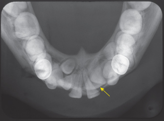
Maxillary and mandibular occlusal radiographs provide useful supplemental information on the position of impacted teeth, especially canines and premolars (White and Pharoah 2004). A maxillary occlusal radiograph is taken as a pretreatment baseline view of the midpalatal suture whenever a rapid maxillary expander is used in treatment. The radiograph can be repeated during the expansion of the arch to observe whether the midpalatal suture has opened and how much it opened. Figure 5.13 is an occlusal view of the patient illustrated in Figure 5.11. The canines are far forward on the palate, near the roots of the permanent incisors. Also note the image of a supernumerary tooth in the middle of the palate between the canines. The supernumerary tooth is a mesiodens that is undergoing resorption, a fate common to many of these teeth. Figure 5.14 shows impacted maxillary second premolars in an adolescent patient. The teeth are erupting toward the midline suture of the palate. Figure 5.15 illustrates the opening of the mid palatal suture of an adolescent patient treated with a rapid maxillary expander. Note the V-shaped opening of the maxillary suture and the diastema created by the appliance. The mandibular occlusal radiograph of an orthodontic patient with cleidocranial dysostosis is shown in Figure 5.16. Note the presence of several supernumerary teeth and the severely impacted mandibular left permanent canine. The impacted canine was located on the labial side of the alveolus and was brought into the arch through orthodontic treatment.
Figure 5.13. View of two impacted canines and a resorbing supernumerary tooth located in the midline of the palate (arrow).
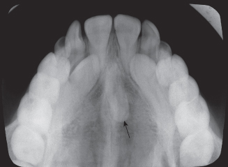
Figure 5.14. View of two ectopic maxillary second premolars.
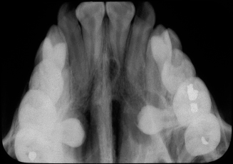
Figure 5.15. View of the midpalatal suture opened by a rapid palatal expander.
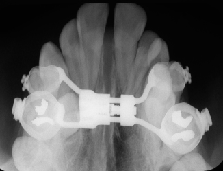
Figure 5.16. View of the mandibular arch of a patient with supernumerary teeth and an impacted mandibular left canine (arrow).
Cone Beam Radiographs
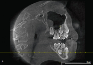
Cone beam radiographs are shown of a patient who had ectopic lower second premolars and a maxillary supernumerary tooth. A conventional panoramic radiograph showed the ectopic teeth through their long axes, not showing where the crowns were located and how long the roots had grown. The supernumerary tooth was located in the upper right palate alongside the canine and premolars. Figure 5.17 is a coronal section through the lower second premolars that gives an excellent view of the developing lower right second premolar and the supernumerary in the upper right palate. Figure 5.18 is a sagittal view of the left mandible illustrating the development of the lower left second premolar. Figure 5.19 is a lingual volumetric view of the lower right second premolar. Figure 5.20 is a lingual volumetric view of the lower left second premolar. Figure 5.21 is an excellent lingual volumetric view of the supernumerary tooth.
Figure 5.17. Coronal view (cone beam computed tomography) of lower second premolars and supernumerary tooth.
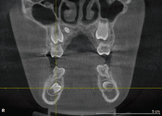
Figure 5.18. Lateral, lingual (cone beam computed tomography) view of the lower left second premolar.
Stay updated, free dental videos. Join our Telegram channel

VIDEdental - Online dental courses


