5
Class II Division 2 Malocclusion
Introduction
A class II division 2 malocclusion is a subdivision of the Angle class II classification and is defined by a class II division 2 incisor relationship, with the incisal edges of the mandibular incisors occluding posterior to the cingulum plateau of the maxillary central incisors, which are retroclined.
Typically, there is an increased and complete overbite stemming from both dento-alveolar and skeletal factors, namely the retroclined labial segments, and reduced Frankfort–mandibular planes angle and lower anterior face height. A traumatic overbite to the gingivae of the lower labial segment labially or the upper incisors palatally is occasionally seen, which in the presence of poor oral hygiene can result in stripping of the gingival attachment.
Skeletally the antero-posterior relationship can range from class I or even a mild class III to severe class II, although the latter is rare and usually associated with a class II division 1 malocclusion. Typically the skeletal pattern is mild class II with a reduced maxillary–mandibular planes angle and a hypo-divergent or brachy-facial form.
The dental arches tend to be short and broad with retroclination of the maxillary central incisors. The lateral incisors are often proclined and rotated mesio-buccally as they escape the control of the lower lip. The lower labial segment can also be retroclined, particularly if the skeletal base relationship is class I or mild skeletal class II, as the lower incisors become trapped behind a retroclined upper labial segment (Mills, 1973). This can result in posterior positioning of B-point compared to pogonion (Fischer-Brandies, 1985). The buccal segment relationship can range from class I to a full unit II, again depending on the severity of the underlying skeletal relationship. Transversely, maxillary buccal crossbites or scissor bites affecting the premolars are common, particularly in more severe low angle skeletal class II cases.
The lips are typically competent, with a high lower lip line, which may rest on the cervical one-third of the maxillary central incisors. The lower lip also covers a greater surface of the upper central incisors as they erupt, leading to retroclination. If, however, the lower lip rests below the lateral incisors, these tend to be proclined. The position of the lower lip is believed to have a significant role in the development of a class II division 2 incisor relationship. If both labial segments are retroclined, there may be little support for the lips in relation to Rickett’s E-line, resulting in a retrusive soft tissue profile. The position of the lower lip in combination with reduced lower face height can also result in a deep labio-mental fold and relative prominence of the chin button.
Class II division 2 malocclusions occur in approximately 10% of Caucasian populations. Classically, hyperactive or hypertonic lips have been implicated in the aetiology of the class II division 2 incisor relationship (Karlsen, 1994). In reality, however, a combination of a reduced lower face height, an increased overbite, a mild skeletal class II base and specific lip position and morphology are usually central to the class II division 2 incisor relationship (McIntyre and Millett, 2006). Indeed, in view of the recognized association of skeletal, dental and soft tissue features, the phrase ‘class II division 2 syndrome’ has been coined. This concept is borne out by the high heritability of class II division 2 malocclusion, complete penetrance being reported in familial studies of monozygotic twins (Markovic, 1992). A reduced crown–root angle has also been reported for the upper incisors in class II division 2 malocclusions, which can contribute to their characteristic inclination (McIntyre and Millett, 2003).
To correct a class II division 2 incisor relationship, overbite reduction is often critical. To promote stability, the inter-incisal angle requires correction by placing the incisal edge of the mandibular incisors anterior to the midpoint or centroid of the upper incisor roots (Houston, 1989). To achieve this, the use of fixed appliances is invariably required to apply torque to the upper labial segment. Space will be required in the upper arch if proclination of the upper incisors is to be prevented during torque delivery. This space can be created by mid-arch extractions or distal movement of the buccal segments.
In a growing patient, the overbite can be reduced effectively using removable appliances with a flat anterior bite plane. Alternatively, in the presence of a skeletal class II pattern, a functional appliance may also be helpful to reduce the overbite. Both of these treatment modalities can allow unimpeded eruption of the molars and premolars. Consequently, the lower face height increases and compensatory growth and adaptation at the condyles stabilizes overbite reduction.
In non-growing patients overbite reduction is more problematic, because without active growth, it is solely reliant on tooth movement. There is usually some scope for proclination of the mandibular incisors, particularly if they are retroclined, which aids overbite reduction and correction of the inter-incisor angle (Mills, 1973; Selwyn-Barnett, 1991). Otherwise, intrusion of the anterior dentition is required to reduce the overbite. True intrusion is mechanically difficult and in severe cases, typically with markedly reduced lower face height, overbite reduction may only be achievable with a combination of fixed appliances and orthognathic surgery.
Case 5.1
| Extra-oral | |
| Skeletal relationship | |
| Antero-posterior | Skeletal class II |
| Vertical | FMPA reduced, lower anterior face height reduced |
| Transverse | No facial asymmetry |
| Soft tissues | Retrusive profile |
| Incompetent lips | |
| Nasio-labial angle: Average | |
| Labio-mental fold: Pronounced | |
| Incisor show | At rest: 2 mm |
| Smiling: 5 mm | |
| Temporo-mandibular joint | Healthy with good range and co-ordination of movement |
| Intra-oral | |
| Teeth present |  |
| Dental health | UL6 and LL6 heavily restored |
| (restorations/caries) | Occlusal caries LR7 |
| Lower arch | Severe crowding with retroclined mandibular incisors |
| Both mandibular canines are disto-angular | |
| LR4 is excluded buccally and the LL5 excluded lingually | |
| Upper arch | Severe crowding with retroclined maxillary incisors |
| Both canines are excluded buccally and distally angulated | |
| Occlusion | Class II division 2 incisor relationship |
| Overjet: 1 mm | |
| Overbite: Increased and complete | |
| Molar relationship: | |
| 1/2 unit class II on the right and class I on the left | |
| Unilateral posterior mandibular buccal crossbite on right side with displacement of the mandible to the right on closure from centric relation (CR) to centric occlusion (CO) | |
| Centre lines: Lower centre line is to the right | |
Summary
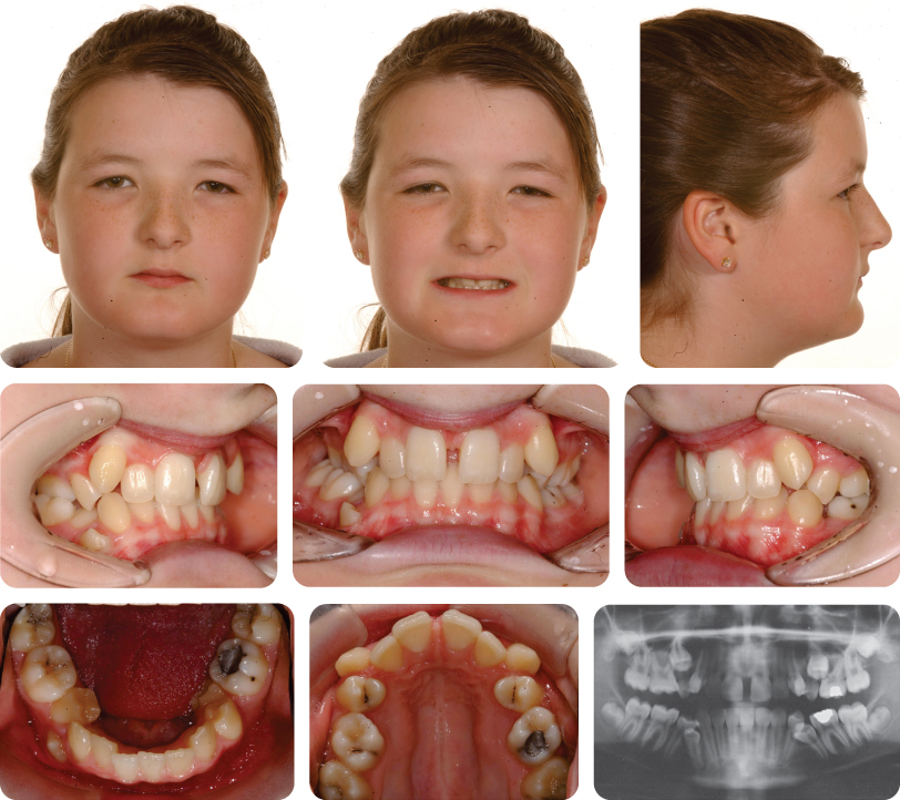
A 13-year-old female presented with a class II division 2 malocclusion on a mild skeletal class II base with reduced vertical dimensions (Figure 5.1). There is severe crowding in both arches, heavily restored first permanent molars on the left hand side and caries in the LR7.
Treatment Plan
- Extraction of the impacted UR5, the UL6, LR4 and LL6
- Upper and lower pre-adjusted edgewise fixed appliances with anchorage re-inforcement
- Retention
What Should Be Done Before Commencing Orthodontic Treatment?
The LR7 should first be restored by the general dental practitioner.
This Is a Case with High Anchorage Demand. What Factors Contribute to This?
- Severe crowding:
- There is severe crowding in the lower arch with impeded eruption of both the LL5 and LR4 due to space deficiency.
- In the maxillary arch the upper buccal segments are severely crowded with both upper second premolars impacted palatally. The upper labial segment is moderately crowded with both canines buccally positioned.
- Canine angulations: All canines are distally angulated. This is especially marked in the upper arch, which will increase the anchorage demands during alignment of the upper labial segment.
- Inclination of the upper labial segment: The upper labial segment is retroclined; this will require torque application to improve the inclination of the teeth.
- Centre line correction is required in both arches.
What Is the Rationale for the Extraction Pattern in this Case?
The LR4 was extracted due to severe crowding and an ectopic position.
The decision to extract the UL6 and the LL6 was based on dental health concerns as both teeth were restored. Removal of these teeth would provide sufficient space to relieve crowding in the buccal segments.
In the maxillary arch, the ectopic unerupted UR5 was extracted due to its unfavourable position.
How Do You Think Anchorage Might Have Been Reinforced in this Case?
In the upper arch, anchorage reinforcement was provided by use of a Nance palatal arch. This was placed to prevent mesial movement of the upper buccal segments. Alternatively, headgear could have been used; given the increased overbite and reduced vertical dimensions, cervical pull headgear would have been the best option.
In the lower arch, anchorage was reinforced by the use of a lingual arch. This facilitated correction of the lower centre line.
Discuss the Mechanics for Centre Line Correction in this Case
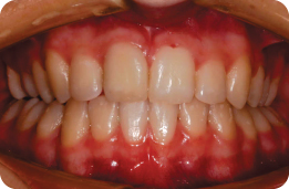
Placement of the lingual arch facilitated lower centre line correction. At the time of appliance placement, a laceback was tied in the lower left quadrant to prevent tip from being expressed in the LL3 bracket. Once in a round 0.018-inch stainless steel archwire centre line, correction was continued by distalizing the LL5 initially, followed by individual retraction of teeth to the left side. The final result is shown in Figure 5.2.
Case 5.2
| Extra-oral | |
| Skeletal relationship | |
| Antero-posterior | Mild skeletal class II |
| Vertical | FMPA: Reduced |
| Lower face height: Reduced | |
| Transverse | Facial symmetry: None |
| Soft tissues | Lip competence: Competent and retrusive to Rickett’s E-line |
| Naso-labial angle: Obtuse and open | |
| Upper incisor show | At rest: 4 mm |
| Smiling: 11 mm | |
| Temporo-mandibular joint | Healthy with good range and co-ordination of movement |
| Intra-oral | |
| Teeth present |  |
| Dental health | Good |
| (restorations, caries) | No active caries and good oral hygiene |
| Lower arch | Mild crowding |
| Incisor inclination: Retroclined | |
| Upper arch | Mild crowding |
| Incisor inclination: Retroclined | |
| Canine position: Left buccal | |
| Right unerupted and not palpable buccally | |
| Occlusion | Incisor relationship: Class II division 2 |
| Overjet: 4 mm to UR2 | |
| Overbite: Increased and complete | |
| Molar relationship: 1/2 unit II bilaterally | |
| Radiographic examination | UR3 was unerupted and ectopic; this tooth was confirmed to be in a palatal position using vertical parallax |
| All third molars present | |
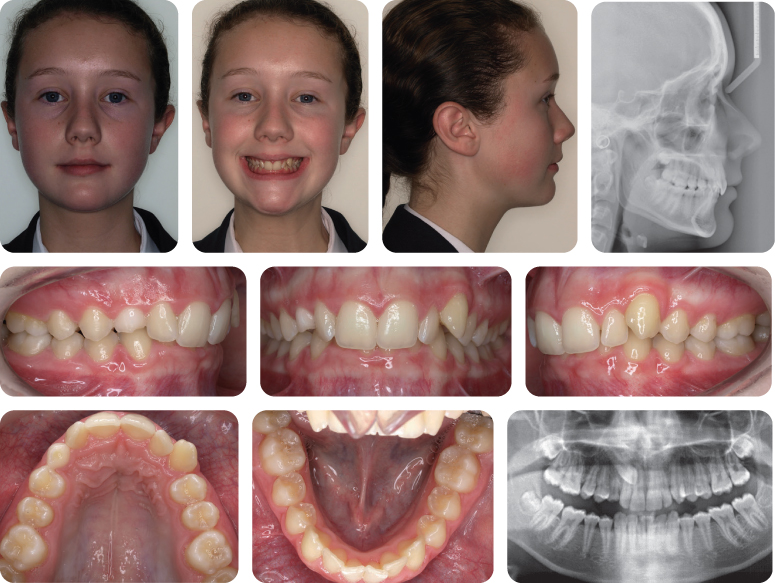
| Cephalometric examination | Confirms the clinical impression of a mild skeletal class II base relationship with reduced FMPA and retroclined upper labial segment (Figure 5.3) |
Summary
A 14-year-old Caucasian female presented with a class II division 2 incisor relationship on a mild skeletal class II base with reduced vertical proportions. The malocclusion was complicated by a palatally impacted UR3, mild crowding of the upper and lower arches, an increased overbite and decreased overjet, and 1/2 unit class II molar relationship bilaterally (Figure 5.3).
Treatment Plan
- Use of headgear and removable appliance to correct the buccal segment relationship to class I while commencing overbite reduction
- Upper and lower pre-adjusted edgewise fixed appliances to level and align arches and correct the incisor relationship
- Exposure and bonding of the UR3 to allow mechanical traction to align UR3
- Long-term retention
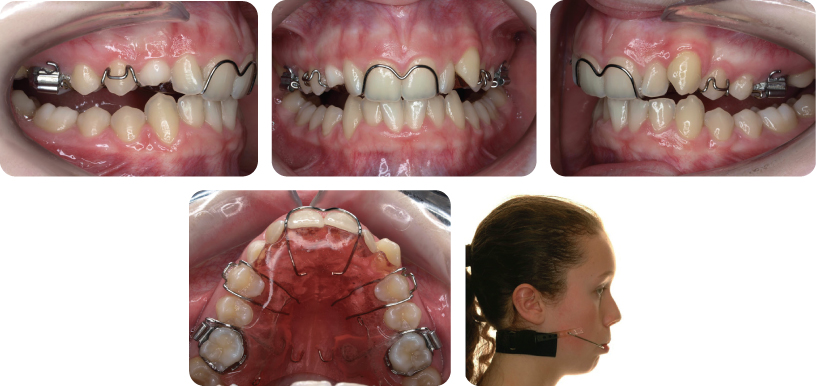
What Treatment Is Being Carried Out in Figure 5.4?
To correct the incisor relationship, the overbite should first be reduced. An effective way of doing this in a growing patient is the use of an anterior bite plane. A removable appliance incorporating a flat anterior bite plane was therefore placed initially.
In addition, space is required in the maxillary arch to correct the buccal segment relationship, torque the upper labial segment and achieve a class I incisor relationship. The removable appliance was supplemented with cervical pull extra-oral traction, directly to bands on the maxillary first molars. The patient was instructed to wear this for 14 hours daily with a 400-g force applied bilaterally. The direction of pull of the headgear creates an extrusive force on the maxillary molars, which aids overbite reduction.
What Are the Principles of Providing a Patient with Headgear?
As well as direction of pull, the other important principles when using headgear are duration of wear, level of force and safety.
For anchorage support when no movement of the molars is desirable, the patient should be instructed to wear the headgear for 10–12 hours a day with a force of between 250 and 350 g applied bilaterally.
To distalize upper molars, as in this case, or to restrict maxillary growth, the force should be increased above 400 g and the headgear worn for at least 14 hours a day. As such, the main limitation of headgear is compliance, as success is reliant on a highly co-operative patient.
There have been a few cases reported in the literature of headgear facebows becoming disengaged from the molar tubes and causing soft tissue injury. The most serious of these is ocular damage with the inner part of the bow penetrating the eye and resulting in infection and blindness. Fortunately, these types of injuries are extremely rare, but given the serious consequences, action should be taken to prevent them. Therefore, at least two safety features should be incorporated in headgear design:
- A locking facebow preventing detachment of the inner bow at night is advocated
- Use of snap-away headgear modules that disengage when excessive force is applied is sensible to prevent catapult injuries.
What Other Features Are Apparent in the Design of the upper removable appliance in Figure 5.4?
The removable appliance also had palatal finger springs placed mesial to the maxillary first molars to aid in their distalization. A Southend clasp on the upper central incisors was also added for retention. The design is a modification of the Acrylic Cervical-Occipital (ACCO) appliance popularized by Cetlin and Ten Hoeve (1983).
What Are the Principles of Overbite Reduction in Class II Division 2 Malocclusions?
Class II division 2 malocclusions require a reduction of the inter-incisal angle, to achieve a class I incisor relationship and stable overbite reduction. A key element in this is the relationship between the lower incisor tip and what is known as the centroid of the upper incisor root (a constructed point half-way along the root). If the lower incisor tip is placed anteriorly to the centroid of the upper incisor, it should lead to greater stability of overbite reduction (Houston, 1989). To achieve this, the maxillary incisor roots require palatal root torque, necessitating fixed appliances and space as retroclined teeth result in a shorter arch length than teeth with appropriate torque. In this case, space was created with headgear and distal movement of the maxillary buccal segments.
What Mechanics Are Being Used in Figure 5.4 to Reduce the Increased Overbite?
There are essentially three ways in which an increased overbite can be reduced:
- Intrusion of the incisors
- Extrusion of the buccal segments
- Proclination of the labial segments.
In this case, the overbite is being reduced initially by extrusion of the buccal segments. This extrusion is achieved primarily using of an upper removable appliance with a flat anterior bite plane. By discluding the posterior teeth, they are free to eru/>
Stay updated, free dental videos. Join our Telegram channel

VIDEdental - Online dental courses


