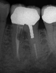
coronal half of each canal had not been obturated. A metal post was present in the distal canal
(Figure 5.3.1).
Figure 5.3.1 Pre-operative radiograph.
Diagnosis and treatment planning
The diagnosis reached was strip perforation of the mesio-lingual canal. The patient was advised that this provisional diagnosis would be confirmed once the crown was removed from the tooth.
What are the diagnostic signs of perforations?
Diagnosis of perforations may be confirmed from clinical signs and radiological findings. Clinically, perforations may exhibit bleeding from the perforation site. Assessment of bleeding can be performed indirectly by inserting paper points into root canals. This is useful for smaller perforations and strip perforations. The level of blood on the paper point will indicate the level of the perforation. The patient may feel a sudden sharp pain during treatment when the perforation occurs. With long-standing perforations, increased probing depths may be detected at the perforation site. Electronic apex locators may also be used to confirm the presence and location of a perforation.
Multiple parallax radiographs, including bitewing radiographs, are necessary for diagnosis. It may be useful to insert a small file (size 10 or 15) into the canal/perforation site to confirm if there is indeed a deviation from the root canal. Radiographs of long-standing perforations may reveal a radiolucency at the site of the perforation. However, perforations on buccal or lingual root surfaces may be difficult to detect due to the overlying anatomical structures. Cone beam computed tomography (CBCT) scans are a useful adjunct to conventional radiographs as they provide three-dimensional views which may aid diagnosis and facilitate treatment planning.
What were the treatment options?
- Non-surgical perforation repair of the 36 without revision of root treatment.
- Root canal re-treatment of the 36 with non-surgical perforation repair.
- Periradicular surgery of the 36 in order to repair the perforation.
- Extraction of the 36, and subsequent restoration of the space with a fixed bridge or an implant retained crown.
Stay updated, free dental videos. Join our Telegram channel
VIDEdental - Online dental courses


