Chapter 4 Head, Neck, and Dental Anatomy
REGIONS OF THE HEAD
Regions of the head include frontal, parietal, occipital, temporal, orbital, nasal, infraorbital, zygomatic, buccal, oral, mental regions. Specific landmarks are noted for each region.
 •
• Oral Cavity

REGIONS OF THE NECK
SKULL
Skull (22 bones), with single and paired bones. Immovable, with EXCEPTION of mandible and temporomandibular joint (TMJ). Has movable articulation with bony vertebral column in neck area. Contains bony openings for important nerves and blood vessels (Table 4-1). Has paranasal sinuses within that serve to lighten bony mass. Has many associated processes that are involved in important structures. To study its landmarks, important to view from superior, lateral, inferior, anterior. Its bones are divided into three categories: cranial, facial, hyoid, which are noted on each view; each category is discussed separately next.
Table 4-1 Bony openings in the skull and their associated nerves and blood vessels
| Bony opening | Bony location | Nerves and vessels |
|---|---|---|
| Carotid canal | Temporal | Internal carotid artery |
| Cribriform plate with foramina | Ethmoid | Olfactory nerves |
| External acoustic meatus | Temporal | (Opening to tympanic cavity) |
| Foramen lacerum | Sphenoid, occipital, temporal | (Cartilage) |
| Foramen magnum | Occipital | Spinal cord, vertebral arteries, eleventh cranial nerve |
| Foramen ovale | Sphenoid | Mandibular division of fifth cranial nerve |
| Foramen rotundum | Sphenoid | Fifth cranial nerve |
| Foramen spinosum | Sphenoid | Middle meningeal artery |
| Greater palatine foramen | Palatine | Greater palatine nerve and vessels |
| Hypoglossal canal | Occipital | Ninth cranial nerve |
| Incisive foramen | Maxilla | Nasopalatine nerve and branches of sphenopalatine artery |
| Inferior orbital fissure | Sphenoid and maxilla | Infraorbital and zygomatic nerves, infraorbital artery, ophthalmic vein |
| Infraorbital foramen and canal | Maxilla | Infraorbital nerve and vessels |
| Internal acoustic meatus | Temporal | Seventh and eighth cranial nerves |
| Jugular foramen | Occipital and temporal | Internal jugular vein and ninth, tenth, eleventh cranial nerves |
| Lesser palatine foramen | Palatine | Lesser palatine nerve and vessels |
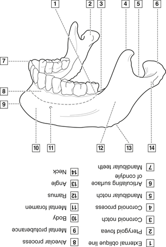
| Mandibular foramen | Mandible | Inferior alveolar nerve and vessels |
| Mental foramen | Mandible | Mental nerve and vessels |
| Optic canal and foramen | Sphenoid | Optic nerve and ophthalmic artery |
| Petrotympanic fissure | Temporal | Chorda tympani nerve |
| Pterygoid canal | Sphenoid | Area nerves and vessels |
| Stylomastoid foramen | Temporal | Seventh cranial nerve |
| Superior orbital fissure | Sphenoid | Third, fourth, sixth cranial nerves and ophthalmic nerve and vein |
From Fehrenbach MJ, Herring SW: Illustrated anatomy of the head and neck, ed 3, St. Louis, 2007, Saunders/Elsevier.
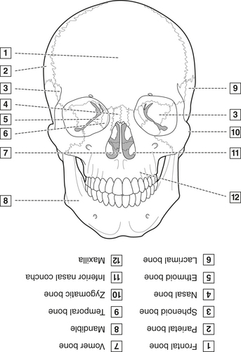
Figure 4-4 Anterior view of skull.
(From Fehrenbach MJ, ed: Dental anatomy coloring book, St. Louis, 2008, Saunders/Elsevier.)
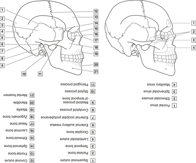
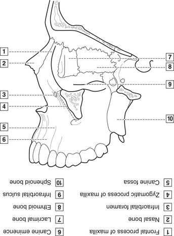
Figure 4-5 Lateral view of skull.
(From Fehrenbach MJ, ed: Dental anatomy coloring book, St. Louis, 2008, Saunders/Elsevier.)
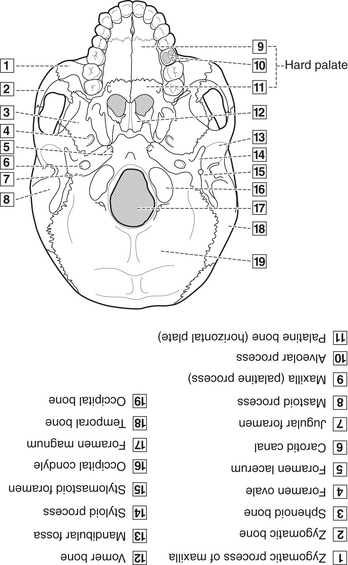
Figure 4-6 Inferior view of external surface of skull.
(From Fehrenbach MJ, ed: Dental anatomy coloring book, St. Louis, 2008, Saunders/Elsevier.)
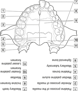
Figure 4-7 Inferior view of hard palate.
(From Fehrenbach MJ, ed: Dental anatomy coloring book, St. Louis, 2008, Saunders/Elsevier.)
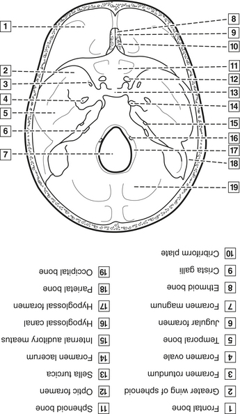
Figure 4-8 Superior view of internal surface of skull.
(From Fehrenbach MJ, ed: Dental anatomy coloring book, St. Louis, 2008, Saunders/Elsevier.)
Cranial Bones
Facial Bones
MUSCLES OF HEAD AND NECK
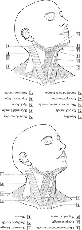
Cervical Muscles
Cervical muscles include SCM and trapezius.
MUSCLES OF FACIAL EXPRESSION
Muscles of facial expression act in various combinations to alter appearance of face; innervated by the facial nerve, seventh (VII) cranial nerve (Figure 4-11).
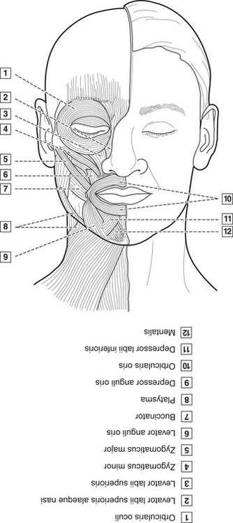
Figure 4-11 Muscles of facial expression.
(From Fehrenbach MJ, ed: Dental anatomy coloring book, St. Louis, 2008, Saunders/Elsevier.)
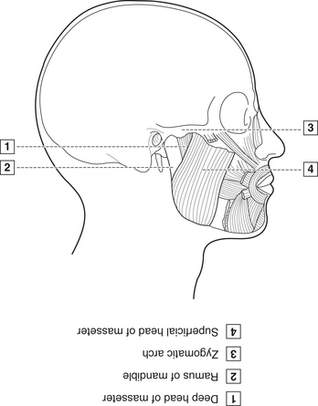
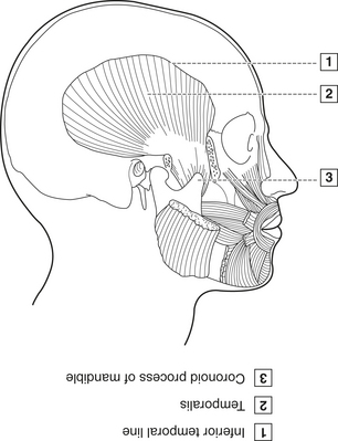
Muscles of Mastication
Muscles of mastication are paired muscles that attach to mandible and work with TMJ to accomplish movements of mandible: depression, elevation, protrusion, retraction, lateral deviation (Figures 4-12 and 4-13). Innervated by mandibular division of fifth (V) cranial nerve (trigeminal). Include masseter, temporalis, medial pterygoid, lateral pterygoid. Involved in temporomandibular joint disorder (TMD). See later discussion on occlusion in regard to masseter.
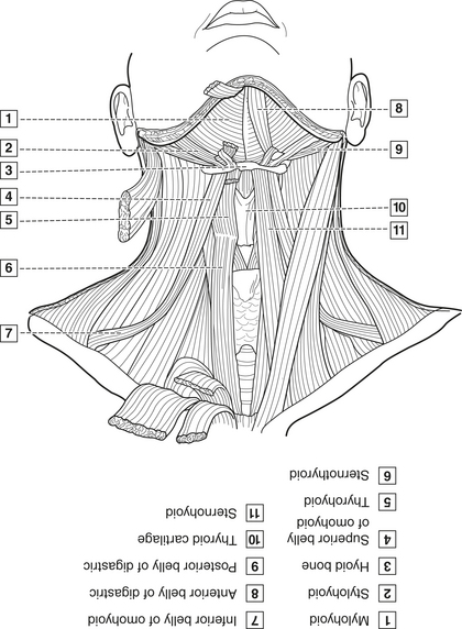
Hyoid Muscles
Hyoid muscles are located superficially in neck tissues, attached to hyoid. Assist in the actions of mastication and swallowing (Figure 4-14).
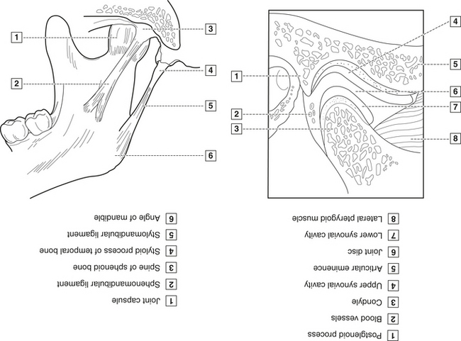
TEMPOROMANDIBULAR JOINT
Temporomandibular joint (TMJ) is located on each side of the head and allows movement of the mandible for speech and mastication (Figure 4-15). Innervated by mandibular division of fifth cranial nerve (trigeminal) and blood supply from the external carotid artery. Patients may experience a temporomandibular joint disorder (TMD) of one or BOTH of these joints. Bones of the TMJ are discussed in skeletal system section.
CLINICAL STUDY
| Age | 58 YRS | SCENARIO |
| Sex |  Male Male  Female Female |
The first afternoon patient has not had a dental examination since her last dentist retired 2 years ago. An initial extraoral examination indicates that her zygomatic region is fi rm and enlarged; she currently has no pain or discomfort in the jaw area. She does admit to frequently grinding her teeth at night. |
| Height | 5′6″ | |
| Weight | 75 LBS | |
| BP | 115/68 | |
| Chief Complaint | “My jaws really ache when I wake up.” | |
| Medical History |
ARTERIAL BLOOD SUPPLY OF THE HEAD AND NECK
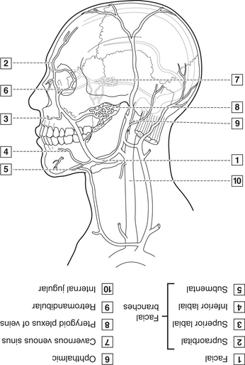
VENOUS DRAINAGE OF THE HEAD AND NECK
Veins are symmetrically located but have greater variability in location than do arteries. Veins anastomose freely and generally are larger and MORE numerous than arteries in same tissue. Internal jugular drains brain and other tissue; external jugular drains only some extracranial tissues, with many anastomoses between them. Internal and external jugular are major venous drainage vessels of head and neck (Figure 4-17). Leaving the head near the base of neck, veins become larger. Vascular system is capable of spreading infection or cancerous cells in head and neck area because valveless veins control direction of blood flow.
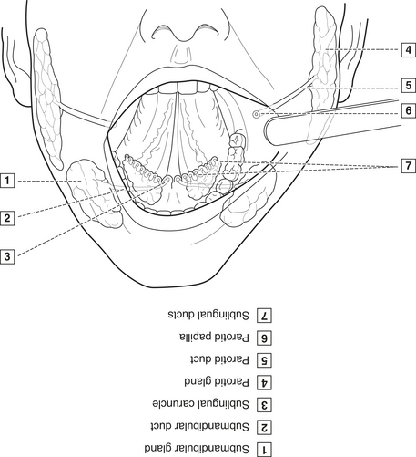
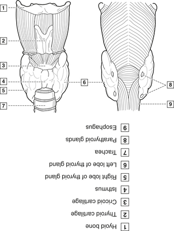
GLANDULAR TISSUE
Glandular tissues include the lacrimal, salivary, thyroid, parathyroid, thymus glands.
CLINICAL STUDY
| Age | 49 YRS | SCENARIO |
| Sex |  Male Male  Female Female |
The intraoral examination of the patient reveals a number of carious lesions, and the oral mucosa appears quite dry. |
| Height | 6′5″ | |
| Weight | 175 LBS | |
| BP | 112/62 | |
| Chief Complaint | “My mouth is so sore when I eat!” | |
| Medical History | Recent radiation therapy for low-grade adenoid cystic carcinoma of the submandibular gland after surgical removal | |
| Current Medications | None | |
| Social History |
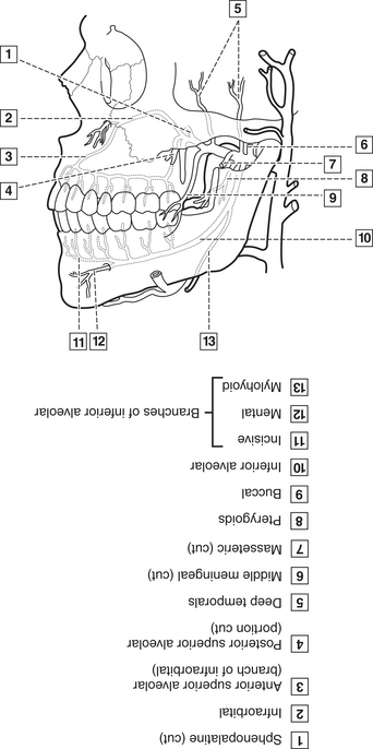
TRIGEMINAL NERVE AND SENSORY ROOT
Trigeminal nerve (fifth cranial nerve [V]) is formed by ophthalmic, maxillary, mandibular branches.
Stay updated, free dental videos. Join our Telegram channel
VIDEdental - Online dental courses


